. 2009 May;83(9):4528-37.
doi: 10.1128/JVI.01910-08. Epub 2009 Feb 11.

Human immunodeficiency virus integrates directly into naive resting CD4+ T cells but enters naive cells less efficiently than memory cells

Affiliations

Human immunodeficiency virus integrates directly into naive resting CD4+ T cells but enters naive cells less efficiently than memory cells

Jihong Dai et al. J Virol. 2009 May.

Abstract

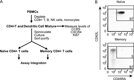
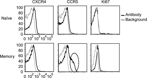
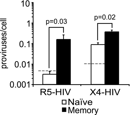
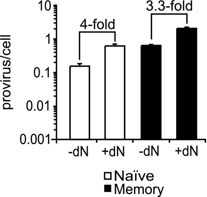
Resting CD4(+) T cells restrict human immunodeficiency virus (HIV) infection at or before reverse transcription, resulting in slower kinetics of reverse transcription. In a previous study, we showed that, despite this restriction at reverse transcription, HIV integration occurs in resting CD4(+) T cells, albeit with slower kinetics. In that study, the resting T cells were a mixture of memory and naïve cells. Here we asked whether the more quiescent naïve cell subset could be directly infected by HIV and, if so, whether the level of integration in naïve cells was comparable to that in memory cells. We found that HIV integrates in the naïve subset of resting CD4(+) T cells without prior activation of the cells. The level of integration (proviruses/cell) in naïve cells was lower than that in memory cells. This difference between naïve and memory cells was observed whether we inoculated the cells with R5 or X4 HIV and could not be explained solely by differences in coreceptor expression. The presence of endogenous dendritic cells did not change the number of proviruses/cell in memory or naïve cells, and deoxynucleoside pools were equally limiting. Our results instead indicate the existence of a novel restriction point in naïve T cells at viral fusion that results in reduced levels of fusion to naïve CD4(+) T cells. We conclude that HIV can integrate into both naïve and memory cells directly. Our data further support our hypothesis that integrated proviral infection of resting T cells can be established without T-cell activation.


Figures

FIG. 1.
FIG. 1.
(A) Outline of purification, infection, and sorting strategy of memory and naïve resting CD4+ T cells. (B) After resting CD4+ T cells were sorted, naïve cells contained 3% contaminating memory cells and memory cells contained 3% contaminating naïve cells. The CD4+ cells were sorted by selecting for CD3+ cells that lacked activation markers (HLA-DR, CD69, and CD25) and had the pattern of expression of CD45RA and CD62L as shown. Fluorescence-minus-one controls were used to place the L-shaped gate that measures the purity of naïve and memory resting cells. The fluorescence-minus-one gates were placed so that 1% of unstained cells appeared as stained. Less than 1% of the cells were endogenous activated (not shown) as defined in our prior papers (2, 62).
FIG. 2.
FIG. 2.
Expression levels of CCR5, CXCR4, and Ki67 in memory and naïve resting CD4+ T cells. The level of expression of the HIV coreceptors CCR5 and CXCR4 and the level of expression of the cellular activation markers Ki67 were assessed by flow cytometry. A subset of memory cells (dashed oval) expressed higher levels of CCR5 than did most memory cells. Minimal expression of the cell cycle marker Ki67 was detected in the memory subset, and no expression was detected in the naïve cells. Unstained cells were used to show the background level for CXCR4 and CCR5 staining. An isotype control was used to determine the background level for the Ki67 intracellular staining.
FIG. 3.
FIG. 3.
X4 HIV integration is detected in naïve resting CD4+ T cells but at lower levels than in memory resting CD4+ T cells. CD4+ T cells were infected by spinoculation with R5 HIV (n = 2 for pRF1 and n = 2 for BaL) or X4 HIV (pNL4-3), cultured for 3 days, and sorted into naïve and memory resting cells. Integration (proviruses/cell) was measured by Alu-PCR. The dashed line represents the expected level of integration from contaminating memory cells (3% contaminants). The bars represent the averages of four independent R5 experiments and five independent X4 experiments. Error bars represent the standard errors. Integration was measured two to four times in each independent experiment. The P values were calculated with the Wilcoxon rank-sum test.
FIG. 4.
FIG. 4.
The presence of endogenous, unstimulated DCs does not influence the susceptibility of resting naïve or memory cells to HIV integration. (A) FACS analysis of spinoculated cells before and after DC depletion. CD4+ T-cell and DC mixtures were exposed to anti-BDCA-1 and anti-BDCA-4 PE-labeled antibodies followed by depletion of BDCA-1+ and BDCA-4+ DCs with anti-PE beads. The cells were then analyzed for the presence of DCs and T cells by staining with anti-BDCA-1, anti-BDCA-4, and anti-CD3 antibodies. (B and C) CD4+ cells depleted or not depleted of endogenous DCs were infected with BaL (R5 HIV) (B) or pNL4-3 (X4 HIV) (C) by spinoculation. At 3 days postinoculation, resting naïve and memory cells were sorted and integration (proviruses/cell) was measured by Alu-PCR. Error bars represent the standard deviations of three to four measurements of integration. The P values were calculated with the Wilcoxon rank-sum test. Similar results were obtained when the DCs were depleted with anti-HLA-DR instead of BDCA-1 and BDCA-4 (not shown).
FIG. 5.
FIG. 5.
Limiting dNs are not responsible for lower susceptibility of naïve resting cells to HIV integration compared to memory resting CD4+ T cells. CD4+ T cells were infected with pNL4-3 (X4 HIV) by spinoculation, followed by culture for 3 days and then sorting into naïve and memory resting CD4+ T cells. The inoculation and culture of the cells were conducted in the presence or absence of 50 μM of dNs. Integration (proviruses/cell) was measured by Alu-PCR. Error bars represent the standard deviations of three measurements of integration.
FIG. 6.
FIG. 6.
Fusion of R5- and X4-tropic HIV to naïve cells is less efficient than that to memory resting CD4+ T cells. CD4+ T-cell and DC mixtures were spinoculated with either pNL-AD8 (R5 HIV) (B) or pNL4-3 (X4 HIV) (D) particles that carry BlaM-Vpr. After inoculation the cells were loaded with the dye CCF2-AM. Uninfected cells (A and C) loaded with CCF2-AM were used as a negative control for each inoculation. After infection of the cells with R5 HIV (B) or X4 HIV (D), naïve and memory resting CD4+ T cells were gated for cells that had taken up the green dye and fusion was measured based on the percentage of cells that fluoresced blue. Fusion was measured independently among the naïve (CD45RA+ CD62L+) and memory (CD45RA and/or CD62L) subsets of resting CD4+ T cells (CD69, CD25, and HLA-DR negative). This experiment is representative of four.
FIG. 7.
FIG. 7.
Both R5 HIV and X4 HIV bind equally to naïve and memory resting CD4+ T cells. Sorted naïve (CD45RA+ CD62L+) and memory (CD45RA and/or CD62L) resting CD4+ T cells were spinoculated with BaL (R5 HIV) and pNL4-3 (X4 HIV) for 2 h at 25°C. The level of viral binding was estimated by measuring the level of cell-associated p24Gag by ELISA immediately after inoculation and after removal of unbound virions. The graph represents the averages of a total of six measurements of viral binding from three independent inoculations. Error bars represent the standard errors. The P values were calculated with the Wilcoxon rank-sum test.
FIG. 8.
FIG. 8.
HIV reverse transcription is less efficient in naïve than in memory resting CD4+ T cells. The efficiencies of integration (proviruses/SST) are similar in both naïve and memory resting CD4+ T cells. CD4+ T-cell and DC mixtures were infected with an R5 HIV (BaL for two experiments and pRF1 for two experiments) or an X4 HIV (pNL4-3) (five experiments) by spinoculation, cultured for 3 days, and then sorted into naïve and memory resting CD4+ T cells. (A) Late reverse transcription was measured by quantitative PCR, using primers that detect the SST of reverse transcription. (B) The ratios of integration to total DNA (proviruses/SST) were obtained by dividing the data in Fig. 3 by the data in panel A. Error bars represent the standard errors of four (R5) and five (X4) independent experiments. The P values were calculated with the Wilcoxon rank-sum test.
FIG. 9.
FIG. 9.
VSV-G-pseudotyped HIV infects both naïve and memory resting CD4+ T cells with similar efficiencies. CD4+ T cells were infected with RF1 (R5 HIV) or pNL4-3 (X4 HIV) particles pseudotyped with VSV-G by spinoculation, followed by culture and sorting into naïve and memory resting CD4+ T cells. Integration was measured as described for Fig. 3 to 5. Error bars represent the standard errors of two independent experiments. The P values were calculated with the Wilcoxon rank-sum test. The X4 HIV(VSV-G) used for these experiments is five times more potent than the R5 HIV(VSV-G) as assessed by the level of reverse transcripts present in CEM-ss cells 24 h after a single-round infection.

References

    1. Adachi, A., H. E. Gendelman, S. Koenig, T. Folks, R. Willey, A. Rabson, and M. A. Martin. 1986. Production of acquired immunodeficiency syndrome-associated retrovirus in human and nonhuman cells transfected with an infectious molecular clone. J. Virol. 59284-291. - PMC - PubMed
    1. Agosto, L. M., J. J. Yu, J. Dai, R. Kaletsky, D. Monie, and U. O'Doherty. 2007. HIV-1 integrates into resting CD4+ T cells even at low inoculums as demonstrated with an improved assay for HIV-1 integration. Virology 36860-72. - PMC - PubMed
    1. Berard, M., and D. F. Tough. 2002. Qualitative differences between naive and memory T cells. Immunology 106127-138. - PMC - PubMed
    1. Berger, E. A., P. M. Murphy, and J. M. Farber. 1999. Chemokine receptors as HIV-1 coreceptors: roles in viral entry, tropism, and disease. Ann. Rev. Immunol. 17657-700. - PubMed
    1. Biancotto, A., J. C. Grivel, S. J. Iglehart, C. Vanpouille, A. Lisco, S. F. Sieg, R. Debernardo, K. Garate, B. Rodriguez, L. B. Margolis, and M. M. Lederman. 2007. Abnormal activation and cytokine spectra in lymph nodes of people chronically infected with HIV-1. Blood 1094272-4279. - PMC - PubMed

Publication types