. 2009 May;83(9):4081-91.
doi: 10.1128/JVI.02591-08. Epub 2009 Feb 11.

Adenovirus induction of IRF3 occurs through a binary trigger targeting Jun N-terminal kinase and TBK1 kinase cascades and type I interferon autocrine signaling

Affiliations

Adenovirus induction of IRF3 occurs through a binary trigger targeting Jun N-terminal kinase and TBK1 kinase cascades and type I interferon autocrine signaling

Marcelo Nociari et al. J Virol. 2009 May.

Abstract

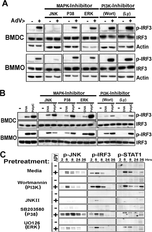
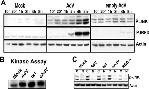
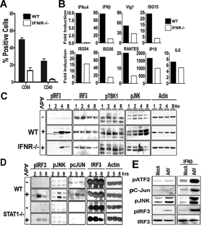
Pathogen recognition is a critical function of immune sentinel cells. Naïve macrophages or dendritic cells (DCs) undergo pathogen-directed activation and maturation, and as mature antigen-presenting cells (APCs), they contribute essential functions to both innate and adaptive immunity. Using recombinant adenovirus (rAdV) as a model for murine APC activation by DNA viruses, we demonstrate a critical role for stress kinase activation in cell intrinsic and extrinsic antiviral signaling cascades. We propose two viral triggers, viral capsid and viral DNA, are required for APC activation. Endosomal escape and presentation of cytosolic rAdV DNA induces phosphorylation of TANK-binding kinase 1 (TBK1) at serine 172 but does not induce IkappaB kinase epsilon activity as determined by in vitro kinase assays. However, induction of TBK1 alone is not sufficient for interferon regulatory factor 3 (IRF3) phosphorylation. We show that capsid-dependent activation of Jun N-terminal kinase (JNK) stress kinase is a necessary step, licensing TBK1 phosphorylation of IRF3 at Ser 396. A second later phase of JNK activity is required to coordinate phosphorylation of JNK-dependent transcription factors (c-Jun/ATF2) with activated IRF3 in the induction of primary IRF3-responsive transcripts. Finally, we demonstrate that maximal JNK/TBK1/IRF3 stimulation by rAdV depends on an intact type I interferon (IFN) signaling cascade. By requiring multiple viral triggers and type I IFN autocrine regulation, APCs have an inherent fail-safe mechanism against inappropriate activation and maturation.


Figures

FIG. 1.
FIG. 1.
TBK1, but not IKKɛ, mediates C-terminal phosphorylation of IRF3 in response to AdV DNA. (A) rAdV induces TBK1 and C-terminal IRF3 phosphorylation with complementary kinetics. Immunoblot of lysates from macrophages treated with vehicle (mock) or rAdV for the indicated times (in hours). Activation of TBK1 and IRF3 was detected with anti-phospho-specific Ser172-pTBK1, and Ser396-pIRF3 antibodies, respectively. Endogenous levels of TBK1 and IRF3 were characterized with polyclonal antibodies against the total proteins. (B) Primary and secondary signaling events elicited by AdV infection. Immunoblot of macrophage lysates harvested 4 h after mock and AdV treatments in the absence or presence of cycloheximide. (C) TBK1 and IKKɛ activities induced by AdV. Protein extracts (600 μg) from macrophages treated for 5 h were subjected to immunoprecipitation (IP) by using antibodies to TBK1 or IKKɛ followed by in vitro kinase assays with GST-IRF3188-427as the substrate. C-IRF3, C-terminal IRF3; WB, Western blotting. (D) AdV internalized genomes, but not capsids, activate TBK1 and IRF3. Western blots showing levels of phospho-TBK1, phospho-IRF3, and corresponding β-actin in macrophage lysates harvested at 5 h posttreatment with vehicle (mock), AdV (psoralen-UV-inactivated Ad2), AdV defective in endosomal escape (ts1) (psoralen UV inactivated), AdV empty capsid (eAdV), RGDmtAdV (RGD), and transfected viral DNA. Representative data from one of two experiments are shown for each panel.
FIG. 2.
FIG. 2.
Roles of the MAPK and PI3K pathways in C-terminal phosphorylation of IRF3 by rAdV. (A) C-terminal phosphorylation of IRF3 induced by AdV infection requires JNK, but not PI3K, signals. Ser396-pIRF3, IRF3, and actin Western blots of lysates from dendritic cells and bone marrow-derived macrophages (BMMO) treated for 5 h with vehicle (−) or AdV (+) in the presence of the indicated inhibitor. The inhibitors were 20 μM SB202190 for p38, 50 μM SP600125 for JNK, ERK for JNK inhibitor II, 50 μM UO126 for 2 μM wortmannin (Wort), and 100 μM LY294002 (Ly). (B) C-terminal phosphorylation of IRF3 induced by transfecting nucleic acids requires PI3K signaling. Ser396-pIRF3, IRF3, and actin immunoblots of lysates from macrophages or DCs treated for 2 h with Lipofectamine (− lanes), Lipofectamine plus adenoviral DNA (DNA lanes), or Lipofectamine plus poly(I·C) (PolyIC lanes) in the presence of the indicated inhibitors. (C) Time course experiment of steady-state levels of Thr183/Tyr185-pJNK, Ser396-pIRF3, and Tyr701-pSTAT1 during AdV infection and effect of signaling inhibitors. Lysates from macrophages treated with vehicle or AdV in the presence of indicated inhibitors were collected at successive time points (in hours) and immunoblotted with anti-Thr183/Tyr185-pJNK, pIRF3, or pSTAT1 phospho-specific antibodies. Representative data from one of two experiments are shown for each panel.
FIG. 3.
FIG. 3.
JNK signaling controls accumulation of C-terminal pIRF3 in a manner that is independent of Ser272 TBK1 phosphorylation. (A) Specificity of JNK and PI3K inhibitors at modulating IRF3 activation. The PI panel shows viability, by propidium iodide (PI) staining, of macrophages after 5-h treatment with vehicle (−) or AdV (+) in the presence of the indicated inhibitors (inhib) (Wort, wortmannin). The Relative DNA panel illustrates viral DNA in 5 h AdV-infected macrophages with the indicated inhibitors. The eGFP panel depicts 36 h transfection efficiency in macrophages treated with indicated inhibitors. eGFP, enhanced green fluorescent protein; MFI, mean fluorescence intensity. The CD86 panel demonstrates the effectiveness of indicated inhibitors at blocking the activation of macrophages by AdV. Macrophage activation was assessed by flow cytometric analysis of CD86 upregulation 36 h after infection. (B) Effect of JNK and PI3K inhibitor on TBK1 activation at 5 h postinfection. Ser396-pIRF3, Ser272-pTBK1 Western blots of lysates from macrophages treated with vehicle (Mock), AdV, Lipofectamine plus viral DNA (DNA), or Lipofectamine plus poly(I·C) (PolyI:C) in the presence of the indicated inhibitors. Representative data from one of two experiments are shown for each panel.
FIG. 4.
FIG. 4.
Immediate-early and early AdV-mediated JNK induction is essential for C-terminal IRF3 phosphorylation and promoter activity, respectively. (A) Effect of immediate-early and early JNK induction on downstream signaling events. JNK activation was selectively blocked in macrophages by adding the JNK inhibitor 2 h before (−2h) or 1 h after AdV infection (+1h). Where indicated, macrophages were pretreated with CHX to enhance JNK activation. 20′, 40′, and 90′, 20, 40, and 90 minutes, respectively; 2 and 5, 2 and 5 hours. (B) Puromycin stress activation of JNK superinduces AdV-mediated C-terminal phosphorylation of IRF3. Macrophages were mock treated or infected with AdV in the presence of media alone or puromycin (Puro) (50 mM). At the indicated times (in hours), whole-cell lysates were prepared, and Western blots were analyzed for JNK signaling (p-JNK and p-cJun) and C-terminal IRF3 activation (pIRF3). Preservation of de novo protein synthesis was monitored using pSTAT1 antibody. (C) Effects of immediate-early and early JNK inhibition on transcriptional induction of IRF3-driven genes (Fold Induction). RT-PCR analysis of macrophages treated for 5 h with vehicle or AdV in the presence of indicated inhibitory conditions (IFN-α4, IFN-β, ISG15, ISG54, ISG56, Vig1, and IP10). Representative data from one of two experiments are shown for each panel.
FIG. 5.
FIG. 5.
Immediate-early JNK activation is triggered by extracellular adenoviral capsid and requires RGD penton motifs. (A) Kinetics of JNK and IRF3 induction following infection with rAdV and empty AdV. Immunoblot analysis of lysates from macrophages treated with vehicle (Mock), rAdV, or empty AdV for the indicated periods of time (from 10 minutes [10′] to 8 hours). (B) rAdV, AdV defective in endosomal escape (ts1), and empty capsid (eAdV) activate JNK. Protein extracts (600 μg) from macrophages treated for 1 h were subjected to immunoprecipitation by using a monoclonal antibody to JNK1 followed by in vitro kinase assays with GST-c-Jun1-89 as the substrate. (C) Western blot analysis of cytosolic (C) and nuclear (N) extracts from macrophages treated for 1 h with vehicle (Mock), rAdV (AdV), AdV defective in endosomal escape (ts1), empty AdV capsid (eAdV), and AdV devoid of penton base RGD motifs (RGD−/−). Representative data from one of two experiments are shown for each panel.
FIG. 6.
FIG. 6.
AdV-mediated IRF3 activation is controlled by type I IFN signaling. (A) AdV-induced surface expression of costimulatory molecules is compromised in IFNR−/− APCs. Macrophages from wild-type (WT) and IFNR−/− mice were treated as previously described and analyzed by flow cytometry for presence of costimulatory molecules CD40 and CD86. (B) IRF3-mediated gene expression is enhanced by type I IFN signaling. Real-time RT-PCR of IRF3-dependent mRNAs isolated from 5-h rAdV infection of WT or IFNR−/− macrophages. IL6, interleukin 6. (C) Type I IFN signaling is required for full JNK activation by AdV. Immunoblot of lysates from macrophages treated with vehicle (−) or AdV (+) for the indicated time points (in hours) were analyzed with anti-phospho-specific (Ser396-pIRF3, Thr183/Tyr185-pJNK, Ser172-pTBK1), anti-IRF3, or anti-β-actin antibodies. (D) Compromised pIRF3, pJNK, and p-cJun peak activation in STAT1−/− macrophages. The results of a time course experiment and Western blot analysis of AdV infection of lysates from WT and STAT1−/− macrophages are shown. (E) Pretreatment with IFN-β augments the JNK/IRF3 response to rAdV. Macrophages were treated for 18 h with 1,000 U/ml IFN-β or vehicle (Mock) followed by rAdV. Lysates harvested 5 h postinfection were characterized by immunoblotting as indicated. Representative data from one of two experiments are shown for each panel.
FIG. 7.
FIG. 7.
Activation of innate immunostimulatory cascades during infection with adenovirus. Interaction of adenovirus capsid with the plasma membrane induces a JNK-mediated stress response (step 1) that via N-terminal phosphorylation or recruitment of scaffold proteins (step 2) would change the IRF3 configuration to unmask its C-terminal end. Simultaneously, the presence of double-stranded DNA in the cytosol (step 3) activates an as-yet-unidentified receptor and adaptor system that results in the induction of the TBK1 enzyme. Activated TBK1 induces the phosphorylation of IRF3 (step 4) on specific serine residues clustered on the now-exposed C-terminal part of the molecule, resulting in its homo- or heterodimerization. These dimers then translocate to the nucleus and in coordination with JNK-activated AP-1 induce the transcription of type I IFN genes (IFN-α4 and IFN-β) (step 5). Secreted IFNs activate the type I IFN receptor (step 6). This interaction leads to Tyk2/JAK activation of STAT1 and STAT2 and formation of the transcription factor complex IFN-stimulated gene factor 3 (ISGF3; a heterotrimer of p-STAT1, p-STAT2, and IRF9). Type I IFN-STAT1 activation leads directly or indirectly to) a positive-feedback amplification loop of the JNK/TBK1/IRF3 cytosolic DNA-sensing response cascade (step 7. The integration of the primary activation cascade (intrinsic) with the autocrine (extrinsic) cascade(s) leads to endpoint maturation of the APCs. AP-1, activator protein-1; ISRE, IFN-stimulated response element.

References

    1. Agalioti, T., S. Lomvardas, B. Parekh, J. Yie, T. Maniatis, and D. Thanos. 2000. Ordered recruitment of chromatin modifying and general transcription factors to the IFN-beta promoter. Cell 103667-678. - PubMed
    1. Andrews, J. M., G. C. Newbound, and M. D. Lairmore. 1997. Transcriptional modulation of viral reporter gene constructs following induction of the cellular stress response. Nucleic Acids Res. 251082-1084. - PMC - PubMed
    1. Basner-Tschakarjan, E., E. Gaffal, M. O'Keeffe, D. Tormo, A. Limmer, H. Wagner, H. Hochrein, and T. Tuting. 2006. Adenovirus efficiently transduces plasmacytoid dendritic cells resulting in TLR9-dependent maturation and IFN-alpha production. J. Gene Med. 81300-1306. - PubMed
    1. Chen, W. C., and L. Huang. 2005. Non-viral vector as vaccine carrier. Adv. Genet. 54315-337. - PubMed
    1. Chu, W., X. Gong, Z. Li, K. Takabayashi, H. Ouyang, Y. Chen, A. Lois, D. J. Chen, G. C. Li, M. Karin, and E. Raz. 2000. DNA-PKcs is required for activation of innate immunity by immunostimulatory DNA. Cell 103909-918. - PubMed

Publication types