Comparative Study
. 2009 Feb 11;29(6):1735-42.
doi: 10.1523/JNEUROSCI.5562-08.2009.

Synaptic overflow of dopamine in the nucleus accumbens arises from neuronal activity in the ventral tegmental area

Affiliations
Comparative Study

Synaptic overflow of dopamine in the nucleus accumbens arises from neuronal activity in the ventral tegmental area

Leslie A Sombers et al. J Neurosci. .

Abstract

Dopamine concentrations fluctuate on a subsecond time scale in the nucleus accumbens (NAc) of awake rats. These transients occur in resting animals, are more frequent following administration of drugs of abuse, and become time-locked to cues predicting reward. Despite their importance in various behaviors, the origin of these signals has not been demonstrated. Here we show that dopamine transients are evoked by neural activity in the ventral tegmental area (VTA), a brain region containing dopaminergic cell bodies. The frequency of naturally occurring dopamine transients in a resting, awake animal was reduced by a local VTA microinfusion of either lidocaine or (+/-)2-amino,5-phosphopentanoic acid (AP-5), an NMDA receptor antagonist that attenuates phasic firing. When dopamine increases were pharmacologically evoked by noncontingent administration of cocaine, intra-VTA infusion of lidocaine or AP-5 significantly diminished this effect. Dopamine transients acquired in response to a cue during intracranial self-stimulation were also attenuated by intra-VTA microinfusion of AP-5, and this was accompanied by an increase in latency to lever press. The results from these three distinct experiments directly demonstrate, for the first time, how neuronal firing of dopamine neurons originating in the VTA translates into synaptic overflow in a key terminal region, the NAc shell.


Figures
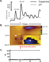
Figure 1.
Figure 1.
Voltammetric recording from an awake rat showing both naturally occurring (indicated with an inverted white triangle) and electrically evoked (arrow) dopamine release, and a pH shift (white asterisk). A, Dopamine concentration changes and pH shift extracted from the voltammetric data using principal component analysis. B, The color plot contains 150 background-subtracted cyclic voltammograms recorded over 15 s. The ordinate is the potential applied to the carbon fiber electrode, the abscissa is time, and the current is depicted in false color. 4 p, Four pulse. C, The residual shows the principal components describe the data well. The dashed line is the threshold for noise predicted by the principal components.
Figure 2.
Figure 2.
Dopamine transients in the NAc shell of an awake rat are suppressed or enhanced by select agents microinfused into the VTA (experiment 1). Representative dopamine signals after microinfusion of saline (left) and after microinfusion of lidocaine (350 nmol, right). Dopamine transients (with a signal-to-noise ratio >5, asterisks) are eliminated after microinfusion of lidocaine. Dopamine concentration changes were extracted from background-subtracted voltammograms.
Figure 3.
Figure 3.
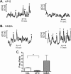
Phasic dopamine signaling is modulated by NMDA receptor-dependent agents in the VTA (experiment 1). A, B, Representative voltammetric data collected in the NAc shell of an awake animal at rest before (left) and immediately after (right) an intra-VTA microinfusion. Dopamine fluctuations are indicated by the asterisks, and are attenuated by microinfusion of AP-5 (A) and enhanced by microinfusion of NMDA (B). Dopamine concentration changes were extracted from the voltammetric data. C, Average dopamine transient frequency ratio (measured over 2 min) before and after microinfusions of saline (n = 6), AP-5 (5 nmol, n = 6, *p < 0.05), and NMDA (0.2 nmol, n = 5).
Figure 4.
Figure 4.
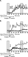
NMDA receptors in the VTA regulate phasic and tonic dopamine in the NAc shell elicited by intravenous cocaine administration (experiment 2). A–C, Each panel contains two superimposed concentration traces collected in a single animal. The animals first received an intra-VTA saline microinfusion during which cocaine was administered intravenously (gray). A second systemic cocaine administration was given 2 h later while saline (n = 5) (A), lidocaine (350 nmol, n = 5) (B), or AP-5 (5 nmol, n = 6) (C) was infused into the VTA (black). Scale bar is the same in all traces.
Figure 5.
Figure 5.
NMDA receptors in the VTA regulate phasic dopamine elicited by reward-predictive cues (experiment 3). A, Representative dopamine concentration trace after microinfusion of saline (left) is attenuated after AP-5 microinfusion (right). B, Average cue-evoked dopamine concentration changes for 50 trials, n = 5 rats. Intra-VTA microinfusion of AP-5, but not saline, significantly decreased the dopamine concentration change elicited by the cue (***p < 0.001).
Figure 6.
Figure 6.
A, Distribution of carbon fiber microelectrode placements in the NAc shell. Coronal diagrams show electrode tip locations for 14 subjects used in this study (black) and for 5 VTA misplacement control subjects (gray). B, Distribution of combination bipolar stimulating electrode/steel guide cannulae placements in the VTA for the same animals. Numbers to the right indicate the anteroposterior coordinates (±0.2 mm) relative to bregma. Coordinates and drawings were taken from a stereotaxic atlas (Paxinos and Watson, 1997).

References

    1. Anderson RM, Fatigati MD, Rompré PP. Estimates of the axonal refractory period of midbrain dopamine neurons: their relevance to brain stimulation reward. Brain Res. 1996;718:83–88. - PubMed
    1. Aragona BJ, Cleaveland NA, Stuber GD, Day JJ, Carelli RM, Wightman RM. Preferential enhancement of dopamine transmission within the nucleus accumbens shell by cocaine is attributable to a direct increase in phasic dopamine release events. J Neurosci. 2008;28:8821–8831. - PMC - PubMed
    1. Arbuthnott GW, Wickens J. Space, time and dopamine. Trends Neurosci. 2007;30:62–69. - PubMed
    1. Britt JP, McGehee DS. Presynaptic opioid and nicotinic receptor modulation of dopamine overflow in the nucleus accumbens. J Neurosci. 2008;28:1672–1681. - PMC - PubMed
    1. Bunney EB, Appel SB, Brodie MS. Electrophysiological effects of cocaethylene, cocaine, and ethanol on dopaminergic neurons of the ventral tegmental area. J Pharmacol Exp Ther. 2001;297:696–703. - PubMed

Publication types

LinkOut - more resources