. 2009 Apr;296(4):C663-71.
doi: 10.1152/ajpcell.00396.2008. Epub 2009 Feb 11.

Membrane targeting and intracellular trafficking of the human sodium-dependent multivitamin transporter in polarized epithelial cells

Affiliations

Membrane targeting and intracellular trafficking of the human sodium-dependent multivitamin transporter in polarized epithelial cells

Veedamali S Subramanian et al. Am J Physiol Cell Physiol. 2009 Apr.

Abstract

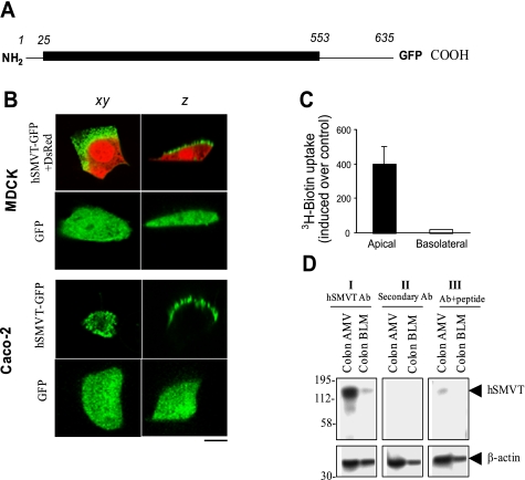
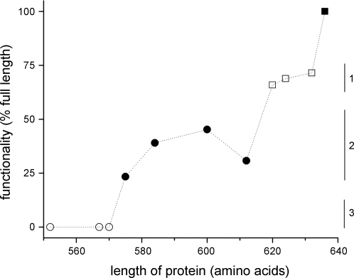
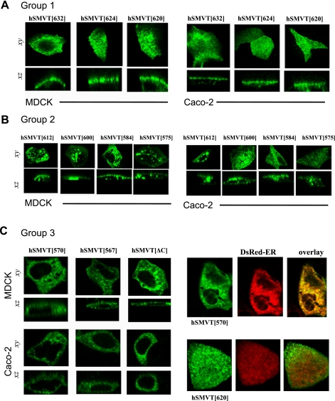
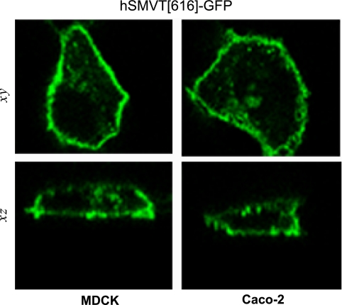
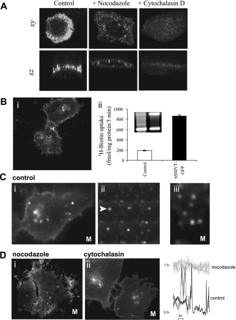
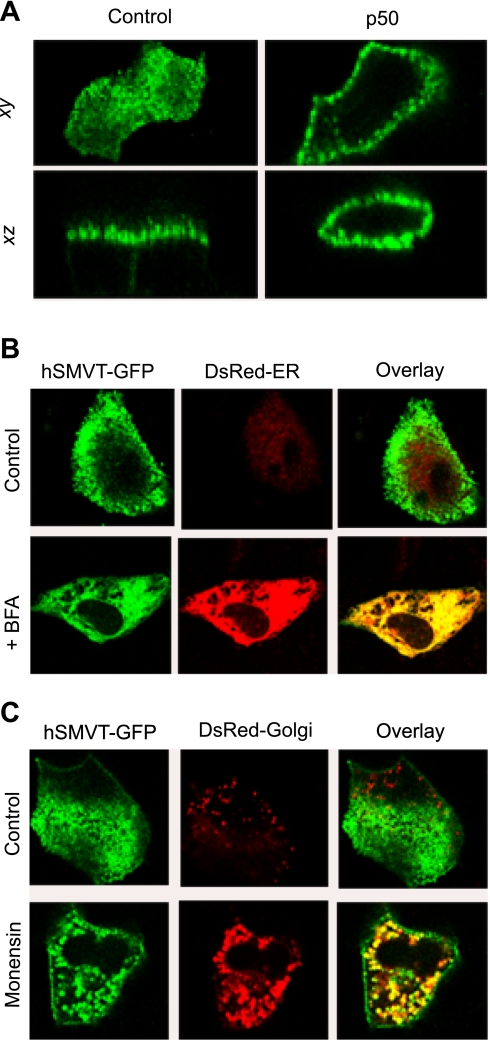
The human sodium-dependent multivitamin transporter (hSMVT) mediates sodium-dependent uptake of biotin in renal and intestinal epithelia. To date, however, there is nothing known about the structure-function relationship or targeting sequences in the hSMVT polypeptide that control its polarized expression within epithelia. Here, we focused on the role of the COOH-terminal tail of hSMVT in the targeting and functionality of this transporter. A full-length hSMVT-green fluorescent protein (GFP) fusion protein was functional and expressed at the apical membrane in renal and intestinal cell lines. Microtubule disrupting agents disrupted the mobility of trafficking vesicles and impaired cell surface delivery of hSMVT, which was also prevented in cells treated with dynamitin (p50), brefeldin, or monensin. Progressive truncation of the COOH-terminal tail impaired the functionality and targeting of the transporter. First, biotin transport decreased by approximately 20-30% on deletion of up to 15 COOH-terminal amino acids of hSMVT, a decrease mimicked solely by deletion of the terminal PDZ motif (TSL). Second, deletions into the COOH-terminal tail (between residues 584-612, containing a region of predicted high surface accessibility) resulted in a further drop in hSMVT transport (to approximately 40% of wild-type). Third, apical targeting was lost on deletion of a helical-prone region between amino acids 570-584. We conclude that the COOH tail of hSMVT contains several determinants important for polarized targeting and biotin transport.


Figures

Fig. 1.
Fig. 1.
Apical targeting of the human sodium-dependent multivitamin transporter (hSMVT)-green fluorescent protein (GFP) fusion protein in renal and intestinal epithelial cells. A: schematic representation of the full-length hSMVT protein (1-635 residues) with GFP fused to the COOH terminus (hSMVT-GFP). B: distribution of hSMVT-GFP and GFP alone in lateral (xy, left) and axial (z, right) section in renal [top; Madin-Darby canine kidney (MDCK)] and intestinal [bottom; human adenocarcinoma (Caco-2)] cell lines. MDCK cells were cotransfected with red fluorescent protein (DsRed) vector to allow resolution of cellular volume. Scale bar is 5 μm. C: [3H]biotin uptake assays in a stable hSMVT-GFP-expressing MDCK cell line grown 5–7 days after confluence on permeable filter supports after introduction of [3H]biotin to the apical (solid) or basolateral (open) chamber. D: expression of the hSMVT protein in native human colon apical membrane vesicles (AMV) and basolateral membrane vesicles (BLM) by Western blotting. All samples were run simultaneously on the same gels (but lanes were grouped for clear presentation), and a representative blot is shown. I: membranes were incubated with primary polyclonal anti-hSMVT antibodies (Ab) raised in rabbits and horseradish peroxidase (HRP)-conjugated secondary antibodies (goat anti-rabbit). II: membranes were incubated with only secondary antibodies. III: anti-hSMVT antibodies pretreated first with antigenic peptide and then incubated with secondary antibodies. Bottom are the same membranes stripped and incubated with human β-actin antibodies. Molecular mass estimations (in kilodaltons) are shown.
Fig. 2.
Fig. 2.
Functionality of hSMVT COOH-terminal tail truncations. Functionality of 10 cytoplasmic tail truncations relative to that of full-length hSMVT-GFP was calculated by measuring the amount of [3H]biotin uptake in a defined period (3 min) corrected for the construct expression level (judged by the mean population fluorescence by flow cytometry). The length of the construct is indicated on the x-axis (amino acids). Data are corralled into 3 groups relative to wild-type (▪, 635 amino acids): ∼75% wild-type functionality (□, group 1), approximately 25–50% wild-type functionality (•, group 2), and transport-null constructs (group 3).
Fig. 3.
Fig. 3.
COOH-terminal truncations disrupt the apical cell surface targeting of hSMVT. A: distribution of indicated hSMVT truncation constructs in MDCK (left) and Caco-2 cells (right) in lateral (xy, top) and axial (xz, bottom) section. All group 1 constructs (Fig. 2) targeted to the apical cell surface. B: group 2 constructs showed progressively impaired apical cell surface targeting in both MDCK (left) and Caco-2 cells (right) shown in lateral (xy, top) and axial (xz, bottom) section. C: left, group 3 constructs were retained intracellularly in both MDCK (left) and Caco-2 cells (right); right, cotransfection of hSMVT[570]-GFP (top) or hSMVT[620]-GFP (bottom) together with DsRed-endoplasmic reticulum (ER) (middle), shown as overlay (right).
Fig. 4.
Fig. 4.
Diverse targeting potential of hSMVT tail truncation. Basolateral targeting of hSMVT[616]-GFP in MDCK (left) and Caco-2 cells (right) shown in lateral (xy, top) and axial (z, bottom) section.
Fig. 5.
Fig. 5.
Effect of cytoskeletal disruption on hSMVT trafficking and targeting. A: MDCK cells treated with nocodazole (20 μM, 30 min; middle) or cytochalasin D (10 μM, 30 min; right) before transient transfection were imaged after 18–24 h and compared with transfected control cells (left). Lateral (xy, top) and axial (xz, bottom) confocal planes are shown. B: i, distribution of hSMVT-GFP trafficking vesicles in a stable human-derived duodenal adenocarcinoma cell line (HuTu-80) cell line grown on cover glass-bottomed petri dishes for 5–7 days; ii, [3H]biotin uptake and RT-PCR of hSMVT mRNA (inset) in the stable HuTu-80 cell line. C: i, total internal reflection fluorescence (TIRF) images of hSMVT vesicular dynamics in HuTu-80 cells (at 37°C; Supplemental Movie S1); ii, montage of single frames (taken at 200-ms intervals) of hSMVT vesicles (e.g., arrow) in control cells; iii, lateral motion of hSMVT containing vesicles (Supplemental Movie S2). D: image stills in cells treated with nocodazole (i; Supplemental Movie S3) and cytochalasin (ii; Supplemental Movie S4). Right: examples of fluorescence profile of vesicles in a nocodazole-treated cell (light gray) to illustrate immobility in the TIRF field relative to time 0 and in a control cell (black) and cytochalasin-treated cell (gray) to illustrate transitions into/out of the TIRF field. M, movie.
Fig. 6.
Fig. 6.
Disruption of hSMVT trafficking. A: effect of dynamitin (p50) overexpression on hSMVT-GFP polarity. Lateral (xy, top) and axial (xz, bottom) confocal images of MDCK cells transfected with hSMVT-GFP (left) or hSMVT-GFP and p50 (right) after 24 h. B: lateral confocal images (xy) showing the effect of brefeldin A (BFA; 5 μg/ml) treatment (14 h, 37°C) on the cellular distribution of hSMVT-GFP in MDCK cells cotransfected with DsRed-ER. C: confocal images (xy) reveal the effect of monensin (5 μM) treatment (14 h, 37°C) on the cellular distribution of hSMVT-GFP in MDCK cells cotransfected with DsRed-Golgi.

References

    1. Anzai N, Miyazaki H, Nohiro R, Khamdang S, Chairoungdua A, Enomoto A, Hirata T, Shin HJ, Sakamoto S, Tomita K, Kanai Y, Endou H. The multivalent PDZ domain-containing protein PDZK1 regulates transport activity of renal urate-anion exchanger URAT1 via its C-terminal. J Biol Chem 279: 45942–45950, 2004. - PubMed
    1. Balamurugan K, Ortiz A, Said HM. Biotin uptake by human intestinal and liver epithelial cells: role of the SMVT system. Am J Physiol Gastrointest Liver Physiol 285: G73–G77, 2003. - PubMed
    1. Balamurugan K, Vaziri ND, Said HM. Biotin uptake by human proximal tubular epithelial cells: cellular and molecular aspects. Am J Physiol Renal Physiol 288: F823–F831, 2005. - PubMed
    1. Banares FF, Lacruz AA, Gine JJ, Esteve M, Gassull MA. Vitamin statues in patients with inflammatory bowel disease. Am J Gastroenterol 84: 744–748, 1989. - PubMed
    1. Bonjour JP Vitamins and alcoholism. Int J Vitam Nutr Res 50: 321–38, 1980. - PubMed

Publication types

MeSH terms

LinkOut - more resources