. 2009 Jun;23(6):1655-62.
doi: 10.1096/fj.08-114611. Epub 2009 Feb 11.

JunD and HIF-1alpha mediate transcriptional activation of angiotensinogen by TGF-beta1 in human lung fibroblasts

Affiliations

JunD and HIF-1alpha mediate transcriptional activation of angiotensinogen by TGF-beta1 in human lung fibroblasts

Amal Abdul-Hafez et al. FASEB J. 2009 Jun.

Abstract

Earlier work showed that TGF-beta1 potently increases angiotensinogen (AGT) gene mRNA in primary human lung fibroblasts. Here the mechanism of TGF-beta1-induced AGT expression was studied in the IMR90 human lung fibroblast cell line. The increase in AGT mRNA induced by TGF-beta1 was completely blocked by actinomycin-D. TGF-beta1 increased the activity of a full-length human AGT promoter-luciferase reporter (AGT-LUC) but did not alter AGT mRNA half-life. Serial deletion analyses revealed that 67% of TGF-beta-inducible AGT-LUC activity resides in a small domain of the AGT core promoter; this domain contains binding sites for hypoxia-inducible factor (HIF)-1 and activation protein-1 (AP-1) transcription factors. TGF-beta1 increased HIF-1alpha protein abundance and the activity of a hypoxia-responsive element reporter; overexpression of HIF-1 increased basal AGT-LUC activity. Both oligonucleotide pulldown and chromatin immunoprecipitation assays revealed increased binding of JunD and HIF-1alpha to the AGT core promoter in response to TGF-beta1. TGF-beta1-inducible AGT-LUC was reduced by an AP-1 dominant negative or by mutation of the AP-1 site. Knockdown of either JunD or HIF-1alpha individually by siRNA partially reduced AGT-LUC. In contrast, simultaneous knockdown of both JunD and HIF-1alpha completely eliminated TGF-beta1-inducible AGT-LUC activity. These data suggest that TGF-beta1 up-regulates AGT transcription in human lung fibroblasts through a mechanism that requires both JunD and HIF-1alpha binding to the AGT core promoter. They also suggest a molecular mechanism linking hypoxia signaling and fibrogenic stimuli in the lungs.


Figures
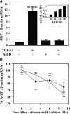
Figure 1.
Figure 1.
TGF-β1 up-regulates AGT gene transcription in IMR90 human lung fibroblasts. A) TGF-β1 (2 ng/ml) was added to IMR90 cells in presence or absence of actinomycin-D (Act-D; 5 μg/ml) for 48 h in serum-free medium. Data represent real-time RT-PCR of AGT mRNA relative to β-actin. Bars are means ± se; n = 3. ***P < 0.001; ANOVA and Student-Newman-Keul’s test. Inset: time-course measurement of AGT/β-actin mRNA post-TGF-β1 treatment (n=2 at each time point). B) TGF-β1 does not increase AGT mRNA stability. IMR90 cells were treated with (open circles) or without (solid circles) TGF-β1 (2 ng/ml) for 48 h, and then actinomycin-D (5 μg/ml) was added for 0, 3, 6, and 9 h, after which total RNA was extracted. Real-time RT-PCR was performed to measure relative levels of AGT and β-actin mRNA and normalized to time 0 (100%). Normalized values are plotted vs. time after actinomycin-D addition to reflect AGT mRNA stability relative to β-actin. Fold difference between controls and TGF-β1 treatment for this experiment is shown in Supplemental Fig. S1. Bars are means ± se; n = 3.
Figure 2.
Figure 2.
AGT core promoter confers TGF-β1 responsiveness in IMR90 cells. A) Deletion analysis of AGT promoter reveals a TGF-β1 response domain close to transcription start site. Full-length (−991 to+22 bp) AGT-LUC firefly luciferase reporter was used to generate the indicated deletion mutants. Each was cotransfected with PRL-CMV Renilla luciferase plasmid into IMR90 cells, followed by exposure to 2 ng/ml TGF-β1 for 24 h, as described in Materials and Methods. Note that most (∼67%) TGF-β-inducible AGT-LUC resides in the domain −46 to +22 bp. Bars are means ± sd; n = 6. *P < 0.05, **P < 0.01, ***P < 0.001 vs. no TGF-β1 for each construct. B) Human AGT promoter proximal sequence (−46 to +3) with known and postulated transcription factor binding domains of interest and known SNPs. TATA box and 2-bp AP-1 mutant (Fig. 3) are indicated; −5/6* denotes true position of G-6A SNP; position of A-20C SNP also is indicated. Numbering discrepancy relative to start codon is due to older SNP nomenclature.
Figure 3.
Figure 3.
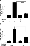
TGF-β1 up-regulates AGT through AP-1. A) A-fos, an AP-1 dominant negative, reduces TGF-β1-induced AGT-LUC. AP-1 dominant negative fos construct (a-fos) was cotransfected into IMR90 cells together with the full-length AGT-LUC construct and PRL-CMV. Transfection was followed by 48 h TGF-β1 (2 ng/ml) treatment followed by dual luciferase assay (see Materials and Methods). B) Mutation of AP-1 site in AGT core promoter reduces TGF-β1-induced AGT-LUC. The full-length AGT-LUC reporter was mutated at −13 and −12 bp in the AP-1 site (see Materials and Methods and Fig. 2B). Wild-type and mutant AGT-LUC constructs were cotransfected with PRL-CMV into IMR90 cells followed by 24 h TGF-β1 (2 ng/ml) treatment. Bars are means ± se. **P < 0.01 (n=3), ***P < 0.001 (n=6); ANOVA and Student-Newman-Keul’s test.
Figure 4.
Figure 4.
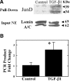
TGF-β1 increases JunD binding to the AGT core promoter. A) Detection of JunD bound to AGT core promoter by Western blotting. A biotinylated oligonucleotide was used in a pulldown method (see Materials and Methods) to identify proteins bound to AGT promoter fragment −46 to +22 bp in untreated (control) or TGF-β1-treated IMR90 cells. Arrows indicate 34- and 39-kDa isoforms of JunD detected by Western blotting, relative to the nuclear proteins lamin A/C, used as loading control for input nuclear extract (NE). B) Quantitative ChIP with antibodies against JunD and specific primers for AGT core promoter fragment −47 to +22. Data are expressed as fold change vs. control. Bars are means ± se; n = 3. *P < 0.05; paired t test.
Figure 5.
Figure 5.
Knockdown of JunD partially reduces TGF-β1-induced AGT transcription in IMR90 cells. A) Gene silencing of JunD in IMR90 cells by siRNA. IMR90 cells were transfected with human JunD siRNA (siJunD), random sequence siRNA (siRan), or mock transfection (mock). After 24 h, JunD was detected by Western blotting of whole cell lysates. Note knockdown of both JunD isoforms but not ∼43-kDa nonspecific protein. B) JunD knockdown reduces the TGF-β1-induced AGT-LUC expression. IMR90 cells were cotransfected with human JunD siRNA (siJunD), random sequence siRNA (siRandom) or mock transfection (mock) together with the full-length AGT-LUC construct and PRL-CMV followed by TGF-β1 (2 ng/ml) for an additional 24 h. AGT-LUC activity was assessed as in Fig. 2. Note partial reduction of AGT-LUC. Bars are means ± se; n = 6. **P < 0.01, ***P < 0.001; ANOVA and Student-Newman-Keul’s test.
Figure 6.
Figure 6.
TGF-β1 increases HIF-1α protein and HIF-1-dependent gene transcription in IMR90 cells. A) TGF-β1 treatment increases nuclear HIF-1α protein. Western blot results for HIF-1α in nuclear extracts of IMR90 cells treated without (control) or with TGF-β1 (2 ng/ml), relative to nuclear proteins lamin A/C used as loading controls. B) Stimulation of hypoxia responsive elements by TGF-β1. IMR90 cells were cotransfected with a hypoxia response element-luciferase reporter (HRE-LUC) and PRL-CMV and were then treated with TGF-β1 for 24 h before dual luciferase assay (see Materials and Methods). Bars are means ± se; n = 6. ***P < 0.001; unpaired t test.
Figure 7.
Figure 7.
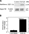
TGF-β1 increases HIF-1α binding to AGT core promoter in IMR90 cells. A) A synthetic biotinylated oligonucleotide of AGT core promoter fragment −46 to +22 was used in pulldown assay with untreated (control) or TGF-β1 treated IMR90 cells followed by HIF-1α detection by Western blotting relative to lamin A/C. B) Real-time PCR quantitation of ChIP DNA using antibodies against HIF-1α and specific primers for AGT core promoter fragment −47 to +32 (see Materials and Methods). Quantitation is expressed in fold change over control. Bars are means ± se; n = 3. ***P < 0.001; unpaired t test.
Figure 8.
Figure 8.
Both HIF-1α and JunD are required for maximal induction of AGT-LUC by TGF-β1 in IMR90 cells. A) Overexpression of HIF-1 increases basal AGT-LUC activity. IMR90 cells were cotransfected with AGT-LUC and PRL-CMV in the presence or absence of two expression plasmids encoding HIF-1, HIF-1α and HIF-β (to ensure sufficient β-partner presence for dimerization with HIF-1α). Cell lysates obtained 24 h post-transfection were used for dual luciferase assay. B) Knockdown of HIF-1α by siRNA. IMR90 cells were treated with TGF-β1 (2 ng/ml) with and without prior exposure to siRNA against HIF-1α (siHIF; see Materials and Methods for details). Note blockade of TGF-β-induced HIF-1 accumulation by the siHIF. C) HIF-1α gene silencing abrogates TGF-β1-induced HRE-LUC expression. IMR90 cells were sequentially transfected with human HIF1α siRNA (siHIF-1α), random sequence siRNA (siRandom), or mock transfection (mock) and 24 h later with HRE-LUC and PRL-CMV. Cells were then treated with TGF-β1 (2 ng/ml) for 24 h and used in dual luciferase assay. D) Both HIF-1α and JunD are required for TGF-β-inducible AGT-LUC activity. IMR90 cells were sequentially transfected with siHIF-1α as in panels B and C or with a mixture of human HIF-1α and JunD siRNA (siHIF-1α+siJunD) and 24 h later, with AGT-LUC and PRL-CMV before TGF-β1 treatment and dual luciferase assay. Note partial elimination of AGT-LUC activity by siHIF-1α alone but complete abrogation by combined knockdown of JunD and HIF-1. See Materials and Methods for details. Bars are means ± se; of n ≥ 6; *P < 0.05, **P < 0.01, *P < 0.001; ANOVA and Student-Newman-Keul’s test. n.s., not significant.

References

    1. Weber K T, Sun Y. Recruitable ACE and tissue repair in the infarcted heart. J Renin Angiotensin Aldosterone Syst. 2000;1:295–303. - PubMed
    1. Yoshiji H, Kuriyama S, Yoshii J, Ikenaka Y, Noguchi R, Nakatani T, Tsujinoue H, Fukui H. Angiotensin-II type 1 receptor interaction is a major regulator for liver fibrosis development in rats. Hepatology. 2001;34:745–7501. - PubMed
    1. Klahr S, Morrissey J J. Comparative study of ACE inhibitors and angiotensin II receptor antagonists in interstitial scarring. Kidney Int Suppl. 1997;63:S111–S114. - PubMed
    1. Ko S H, Hong O K, Kim J W, Ahn Y B, Song K H, Cha B Y, Son H Y, Kim M J, Jeong I K, Yoon K H. High glucose increases extracellular matrix production in pancreatic stellate cells by activating the renin-angiotensin system. J Cell Biochem. 2006;98:343–355. - PubMed
    1. Marshall R P, Gohlke P, Chambers R C, Howell D C, Bottoms S E, Unger T, McAnulty R J, Laurent G J. Angiotensin II and the fibroproliferative response to acute lung injury. Am J Physiol Lung Cell Mol Physiol. 2004;286:L156–L164. - PubMed

Publication types

MeSH terms