Review
. 2009;85(2):55-68.
doi: 10.2183/pjab.85.55.

Evolution of non-coding regulatory sequences involved in the developmental process: reflection of differential employment of paralogous genes as highlighted by Sox2 and group B1 Sox genes

Affiliations
Review

Evolution of non-coding regulatory sequences involved in the developmental process: reflection of differential employment of paralogous genes as highlighted by Sox2 and group B1 Sox genes

Yusuke Kamachi et al. Proc Jpn Acad Ser B Phys Biol Sci. 2009.

Abstract

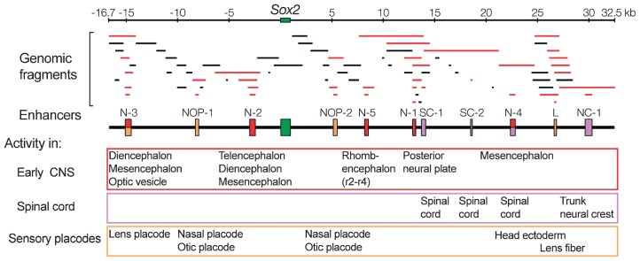
In higher vertebrates, the expression of Sox2, a group B1 Sox gene, is the hallmark of neural primordial cell state during the developmental processes from embryo to adult. Sox2 is regulated by the combined action of many enhancers with distinct spatio-temporal specificities. DNA sequences for these enhancers are conserved in a wide range of vertebrate species, corresponding to a majority of highly conserved non-coding sequences surrounding the Sox2 gene, corroborating the notion that the conservation of non-coding sequences mirrors their functional importance. Among the Sox2 enhancers, N-1 and N-2 are activated the earliest in embryogenesis and regulate Sox2 in posterior and anterior neural plates, respectively. These enhancers differ in their evolutionary history: the sequence and activity of enhancer N-2 is conserved in all vertebrate species, while enhancer N-1 is fully conserved only in amniotes. In teleost embryos, Sox19a/b play the major pan-neural role among the group B1 Sox paralogues, while strong Sox2 expression is limited to the anterior neural plate, reflecting the absence of posterior CNS-dedicated enhancers, including N-1. In Xenopus, neurally expressed SoxD is the orthologue of Sox19, but Sox3 appears to dominate other B1 paralogues. In amniotes, however, Sox19 has lost its group B1 Sox function and transforms into group G Sox15 (neofunctionalization), and Sox2 assumes the dominant position by gaining enhancer N-1 and other enhancers for posterior CNS. Thus, the gain and loss of specific enhancer elements during the evolutionary process reflects the change in functional assignment of particular paralogous genes, while overall regulatory functions attributed to the gene family are maintained.


Figures

Fig. 1
Fig. 1
Expression of Sox2 in chicken embryo at various developmental stages marking neural and sensory primordia, as indicated by in situ hybridization. Anterior is toward the top. Photographs were taken at the same scale. The position of organizer (Hensen’s node) is indicated by an arrowhead. Head ectoderm (E), lens placode (L) and otic vesicle (O) are indicated by arrows. Adapted from Fig. 1A in Uchikawa et al. (2003). Functional analysis of chicken Sox2 enhancers highlights an array of diverse regulatory elements that are conserved in mammals. Dev. Cell 4, 509–519, with permission from Elsevier.
Fig. 2
Fig. 2
Determination of Sox2 enhancers with activities in various and distinct domains of embryonic CNS and sensory placodes. Genomic fragments covering the 50 kb region of chicken Sox2 locus were individually tested for enhancer activity in electroporated chicken embryos. DNA fragments that demonstrated an enhancer activity are shown in red, and functionally determined enhancers are indicated by boxes on the map (middle). Adapted from Fig. 2 in Uchikawa et al. (2003). Functional analysis of chicken Sox2 enhancers highlights an array of diverse regulatory elements that are conserved in mammals. Dev. Cell 4, 509–519, with permission from Elsevier.
Fig. 3
Fig. 3
Conserved activity of enhancers N-1 to N-5 between chicken and mouse. (A) Domains of the embryonic CNS where chicken enhancers N-1 to N-5 show activity in electroporated embryo compared to Sox2 expression (in situ hybridization) of stage 11 embryo. Fluorescence images (green) representing enhancer activities are overlaid on darkened bright-field images to indicate respective embryonic domains. “r” indicates the rhombencephalic domain. The orange speckles in the enhancer N-5 specimen are due to DsRed fluorescence that was derived from co-electroporated control vector and not removed completely by optical filtration. (B) Alignment of chicken and mouse Sox2 enhancers with nucleotide sequence identity expressed as a percentage. (C) Activity of mouse enhancers at E9–9.5 in transgenic mouse embryos, compared to Sox2 in situ hybridization. Transgenes for Sox2 enhancers, except for enhancer N-1, were constructed by introducing each tetrameric Sox2 enhancer of mouse upstream of the hsp68 promoter-LacZ cassette, and primary transgenic embryos were examined. Tetrameric N-1 enhancer in a tk-LacZ vector was used to generate transgenic lines.
Fig. 4
Fig. 4
Correspondence of the functionally defined Sox2 enhancers with blocks of highly conserved sequences found by comparison of chicken and mammalian Sox2 locus sequences. (A) Conserved sequence blocks found in the Sox2 locus sequences of amniotes. Blocks of sequences that show > 60% identity over the stretch of 100 base pairs are indicated by boxes. The 25 sequence blocks No. 1 to No. 25 conserved between chicken and mammalian sequences are indicated on the top. Blocks No. 3, 21 and 25 marked by asterisks are not conserved in the mouse sequence. Sequences conserved between human and mouse genomic sequences but not strongly conserved in the chicken sequence are indicated in blue. (B) Dot matrices comparing DNA sequences of the three animal sequences encompassing enhancer N-5 (conserved sequence block No. 14). A dot indicates a 10 bp sequence with > 60% matching. Between the chicken and mammalian sequences, only the enhancer sequence is significantly conserved, however between human and mouse, the enhancer sequence is embedded in a broader region of possibly non-functional sequence conservation. Adapted from Fig. 6 in Uchikawa et al. (2003). Functional analysis of chicken Sox2 enhancers highlights an array of diversity regulatory elements that are conserved in mammals. Dev. Cell 4, 509–519, with permission from Elsevier.
Fig. 5
Fig. 5
Distinct characteristics of enhancers N-2 and N-1 that regulate Sox2 in anterior and posterior neural plates, respectively. A. In early stage chicken embryos, N-2 and N-1 are the only active Sox2 enhancers, covering un-overlapping anterior (N-2, red fluorescence of mRFP1) and posterior domains (N-1, green fluorescence of EGFP) of the developing CNS. B. Regulatory modules of enhancer N-1 core sequence determined by Takemoto et al. C. When a tk-mCherry vector carrying chicken enhancer N-2 was injected into zebrafish embryo, it was regulated to have activity in the anterior neural plate, whereas analogous tk-Venus vector carrying dimeric chicken N-1 was not activated with regional restriction.
Fig. 6
Fig. 6
Differential range of phylogenetic conservation of Sox2 enhancers. A. Comparison of highly conserved sequence blocks (boxes) and enhancers functionally assessed in higher vertebrates (colored boxes) among six animal species, human, mouse, opossum, chicken, Xenopus and zebrafish. Enhancers showing activity in the brain-forming anterior CNS are connected by red lines, while those in the posterior CNS are connected by blue lines. Most of the enhancers showing activity in the posterior CNS are conserved in amniotes, however conservation is limited in Xenopus and absent in fish. B. Alignment of enhancer N-1 sequences between five animal species. Although the entire sequence is highly conserved among amniotes, sequence conservation is limited to the core-proximal sequences in Xenopus; even in the core sequence only one Lef1 binding sequence among three essential functional elements is conserved in the Xenopus sequence.
Fig. 7
Fig. 7
Expression of group B1 Sox genes in early stage zebrafish embryos. A. RT-PCR analysis of transcripts of Sox1a, Sox1b, Sox2, Sox3, Sox19a and Sox19b in embryos at various stages. β-actin was used as control for the reaction. B. Expression pattern of the genes at various developmental stages indicated by in situ hybridization (blue). Hybridization with no tail probe (orange) was used to mark mesodermal precursors in gastrulating embryos. Developmental fates of early embryonic stages are illustrated for comparison: NNE, non-neural ectoderm; NE, neural ectoderm; D, dorsal; V, ventral; F, forebrain; M, midbrain; H, hindbrain; SC, spinal cord. Arrowheads indicate the site of shield. In 12-somite stage embryos, optic vesicle (ov), otic placode (otp), fore-midbrain boundary (fmb) and mid-hindbrain boundary (mhb) are indicated. Reprinted from Figs. 3, 4 and 6A in Okuda et al. (2006). Comparative genomic and expression analysis of group B1 sox genes in zebrafish indicates their diversification during vertebrate evolution. Dev. Dyn. 235, 811–825, with permission from Wiley-Blackwell.
Fig. 8
Fig. 8
Evolutionary history of genesis of group B1 Sox paralogues as a consequence of multiple rounds of genomic duplication. “Vertebrate group B Sox prototypic composition” is hypothetical. Linkages of “Sox14 and Sox2” and “Sox21 and Sox1” are conserved in human and other primates, cow and chicken, but disrupted in mouse after extensive chromosomal rearrangements (Ensembl 50, July 2008; http://www.ensembl.org/index.html). Linkage of “Sox21 and Sox1” is found in a broader range of vertebrate species, e.g., dog, opossum, and medaka and other fish species, although this is disrupted in zebrafish. Linkages of “Sox14 and Sox2” is reported for platypus. Linkages of these genes in Xenopus genome have not been confirmed.
Fig. 9
Fig. 9
Evolutionary shift of the major “pan-neural” group B1 Sox genes and of posterior coverage of the CNS by Sox2, Sox3 and Sox19 paralogues, as indicated by expression patterns in embryos at comparable developmental stages. (A) Expression pattern in zebrafish embryo of 3 somite stage. Sox19a is the most prevalent, while strong Sox2 expression is mostly confined to the anterior CNS, as in other stages (Fig. 7). Reprinted from Fig. 5B in Okuda et al. (2006). Comparative genomic and expression analysis of group B1 sox genes in zebrafish indicates their diversification during vertebrate evolution. Dev. Dyn. 235, 811–825, with permission from Wiley-Blackwell. Reported Sox3 expression pattern in medaka embryo is analogous to zebrafish. (B) Expression pattern in Xenopus embryos at stage 13 and later, where Sox3 is the major Sox gene expressed. Expression patterns of Sox2 and Sox3 are reproduced from Fig. 1 of Schlosser and Ahrens (2004). Molecular anatomy of placode development in Xenopus laevis. Dev. Biol. 271, 439–466, and that of SoxD is reproduced from Fig. 2 of Mizuseki et al. (1998). SoxD: an essential mediator of induction of anterior neural tissues in Xenopus embryos. Neuron 21, 77–85, with permission from the authors and Elsevier. (C) Expression pattern in chicken embryo at stage 8, where Sox2 expression is dominating. In the chicken, Sox19-paralogous gene has not been identified. Arrows indicate the shift of major group B1 Sox among the species. A and P indicate anterior and posterior, respectively.

References

    1. Hardison R. C. (2000) Conserved noncoding sequences are reliable guides to regulatory elements. Trends Genet. 16, 369–372 - PubMed
    1. Levy S., Hannenhalli S., Workman C. (2001) Enrichment of regulatory signals in conserved non-coding genomic sequence. Bioinformatics 17, 871–877 - PubMed
    1. Uchikawa M., Ishida Y., Takemoto T., Kamachi Y., Kondoh H. (2003) Functional analysis of chicken Sox2 enhancers highlights an array of diverse regulatory elements that are conserved in mammals. Dev. Cell 4, 509–519 - PubMed
    1. Uchikawa M., Takemoto T., Kamachi Y., Kondoh H. (2004) Efficient identification of regulatory sequences in the chicken genome by a powerful combination of embryo electroporation and genome comparison. Mech. Dev. 121, 1145–1158 - PubMed
    1. Woolfe A., Goodson M., Goode D. K., Snell P., McEwen G. K., Vavouri T., et al. (2005) Highly conserved non-coding sequences are associated with vertebrate development. PLoS Biol. 3, e7. - PMC - PubMed

Publication types

MeSH terms

Substances