. 2009;4(2):e4459.
doi: 10.1371/journal.pone.0004459. Epub 2009 Feb 12.

The SmpB-tmRNA tagging system plays important roles in Streptomyces coelicolor growth and development

Affiliations

The SmpB-tmRNA tagging system plays important roles in Streptomyces coelicolor growth and development

Chunzhong Yang et al. PLoS One. 2009.

Abstract

The ssrA gene encodes tmRNA that, together with a specialized tmRNA-binding protein, SmpB, forms part of a ribonucleoprotein complex, provides a template for the resumption of translation elongation, subsequent termination and recycling of stalled ribosomes. In addition, the mRNA-like domain of tmRNA encodes a peptide that tags polypeptides derived from stalled ribosomes for degradation. Streptomyces are unique bacteria that undergo a developmental cycle culminating at sporulation that is at least partly controlled at the level of translation elongation by the abundance of a rare tRNA that decodes UUA codons found in a relatively small number of open reading frames prompting us to examine the role of tmRNA in S. coelicolor. Using a temperature sensitive replicon, we found that the ssrA gene could be disrupted only in cells with an extra-copy wild type gene but not in wild type cells or cells with an extra-copy mutant tmRNA (tmRNA(DD)) encoding a degradation-resistant tag. A cosmid-based gene replacement method that does not include a high temperature step enabled us to disrupt both the ssrA and smpB genes separately and at the same time suggesting that the tmRNA tagging system may be required for cell survival under high temperature. Indeed, mutant cells show growth and sporulation defects at high temperature and under optimal culture conditions. Interestingly, even though these defects can be completely restored by wild type genes, the DeltassrA strain was only partially corrected by tmRNA(DD). In addition, wildtype tmRNA can restore the hygromycin-resistance to DeltassrA cells while tmRNA(DD) failed to do so suggesting that degradation of aberrant peptides is important for antibiotic resistance. Overall, these results suggest that the tmRNA tagging system plays important roles during Streptomyces growth and sporulation under both normal and stress conditions.


Conflict of interest statement

Competing Interests: The authors have declared that no competing interests exist.

Figures

Figure 1
Figure 1. Construction of ssrA-DD mutant of S. coelicolor ssrA gene and detection of tagged proteins.
A. Detail of the sequence and proposed secondary structure of the tag-coding sequence of S. coelicolor tmRNA. Bases in red in ssrA-DD were altered by mutagenesis. B. Northern blot analysis of total RNA isolated from empty vector transformed cells (C = control), and cells expressing tmRNA from a single-copy (S) and multi-copy (M) plasmid carrying ssrA-DD. Ethidium bromide stained ribosomal RNA (rRNA) is shown as a loading control. Note that the probe is generated from full-length ssrA DNA and does not distinguish between the wild type and mutant tmRNA. C. Western blot detection of tmRNADD tagging in S. coelicolor. Total proteins from strains analyzed in panel B were separated by SDS-PAGE and either stained with Coomassie blue (CB) or transferred to PVDF membranes and probed with affinity-purified anti-ssrA-DD tag antibody (WB). Immunocomplexes were detected by ECL. D. Tagging in S. lividans analyzed as in panel C.
Figure 2
Figure 2. Effect of bldA expression on tmRNA-mediated tagging.
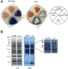
A. Developmental phenotypes were determined for wild type (wt) and ΔbldA strains either transformed with a high copy-number vector with no insert (+vec) or with the same vector containing ssrA-DD (+ssrA-DD). Plates were scanned from the top and bottom to illustrate both spore and pigment production. B. Western blot analysis of the strains carrying ssrA-DD described in A was performed as described in the legend to Figure 1. Arrows highlight tagged bands that are more readily detected in the ΔbldA cells in the magnified image.
Figure 3
Figure 3. Insertional mutagenesis using a temperature-sensitive replicon.
A. Northern blot analysis. Total RNAs were extracted from S. coelicolor strains with marker only, wild type ssrA or ssrA-DD inserted at the attB locus and analysed by Northern blot using a probe that does not distinguish between wild type and mutant tmRNA. Ethidium bromide stained ribosomal RNA (rRNA) is shown as a loading control. B. Schematic diagrams illustrating the anticipated genomic maps of the ssrA locus, the attB locus following integration of ssrA or ssrA-DD, and the ssrA locus following correct integration of the HygR cassette. The expected sizes of NcoI restriction fragments are indicated in each case. Lower case letters to the right of each fragment indicates fragment labeled in panel C. C. Southern blot analysis of NcoI-digested genomic DNA isolated from “positive” strains that were resistant to hygromycin and sensitive to thiostrepton. Ten isolates from each group were analyzed, five of which are illustrated. The top Southern blot was probed with an ssrA probe while the bottom blot was hybridized with a probe for the HygR cassette after the same blot was stripped. Control lane (Con) is genomic DNA from an untransformed wild type strain. Arrows and letters indicate genomic fragments illustrated in Panel B. D. Two of the ten positives from the ssrA/- group with an unusual Southern blot result (one of these is indicated by asterisk in panel B) were further analyzed by genomic PCR of the ssrA region with genomic DNA template from wild type cells used as a control. An ethidium bromide-stained agarose gel is illustrated.
Figure 4
Figure 4. Southern blot analysis of ΔsmpB, ΔssrA and ΔsmpB/ssrA strains.
A. Schematic diagrams of the genomic map of the wild type genomic smpB/ssrA locus and the anticipated structures following integration of the apramycin resistance cassette. The expected sizes of NcoI restriction fragments are indicated in each case. B. Southern blot analysis of NcoI-digested genomic DNA isolated from two independent isolates that were resistant to apramycin and sensitive to kanamycin. After detection with a probe for the aprR cassette (top), the blot was stripped and redetected with an ssrA probe (bottom).
Figure 5
Figure 5. Northern blot analysis of the mutant strains.
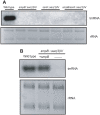
A. Total RNAs from the same isolates shown in Figure 4 were prepared and probed for tmRNA expression by Northern blot. An ethidium bromide-stained agarose gel showing rRNAs is illustrated as loading control. B. The ΔsmpB strain was transformed with an SmpB expression plasmid was compared by Northern blot analysis with control (wild type) and mutant cells without complementation.
Figure 6
Figure 6. Liquid growth phenotypes of the mutant strains.
A. S. coelicolor wild type (left) and ΔssrA mutant (right) strains were grown in YEME medium at 30°C from spores. Phase-contrast micrographs were taken with a Zeiss microscope at 12 h, 48 h, and 72 h. B. Wild type and mutant strains were first grown up from spores in YEME medium in a 30°C shaker at 250 rpm until culture reached log phase (OD 450 = 1–2) and then ground manually to break up mycelium, diluted into fresh YEME medium to equal density of OD 450 at 0.05 and further cultured with shaking at 30°C.
Figure 7
Figure 7. Agar growth and sporulation phenotypes of the mutant strains.
Equal number (based on OD 450) of wild type and ssrA mutant spores were resuspended in 50 µL YEME medium, streaked onto the surface of R2YE plate and incubated at 30°C. At indicated time points, plates were scanned from both top and bottom of the plate to show surface growth and pigment production.
Figure 8
Figure 8. Phenotype at high temperature and mutant complementation.
A. Wild type and mutant spores were plated as previously described in the legend to Figure 7 and grown at 39°C. At day 5 and 14, pictures were taken from the top to show growth and sporulation. B. Wild type and two independent isolates each of the indicated single and double mutants were first cultured in liquid YEME medium to log phase and equal number of cells (based on OD 450) were plated onto R2YE agar plate and grown at 39°C for 5 d. Plates were scanned from top. C. Spores of the ΔssrA strain, both uncomplemented and complemented with ssrA or ssrA-DD, were cultured in YEME in a 250 rpm shaker at 30°C for 3 d and equal densities (based on OD 450) were plated onto R2YE agar plate and further incubated at 39°C for 5 d. D. Spores of ΔsmpB and ΔssrA strains with and without complementing genes were inoculated into YEME medium to an OD 450 of 0.03 and grown at 39°C for 5 d. OD 450 was measured and used to plot the graph. E. Wild type and ΔssrA strains were transformed with a multicopy ssrA-DD plasmid and tagging was analyzed by Western blot as described in Figure 1C legend.
Figure 9
Figure 9. Complementation of hygromycin sensitivity.
Left panel. Wild type and mutant spores (two independent isolates each) were serially diluted 5-fold and plated onto R2YE agar without any drug (upper) or with 5 µg mL−1 hygromycin (lower). Right panel. Spores of mutant cells with and without complementing genes were serially diluted and plated onto R2YE agar plate without drug (upper) or with 5 µg mL−1 hygromycin (lower). All plates were scanned from top after grown at 30°C for 5 d.

Similar articles

Cited by

References

    1. Karzai AW, Roche ED, Sauer RT. The SsrA-SmpB system for protein tagging, directed degradation and ribosome rescue. Nat Struct Biol. 2000;7:449–455. - PubMed
    1. Withey JH, Friedman DI. A salvage pathway for protein structures: tmRNA and trans-translation. Annu Rev Microbiol. 2003;57:101–123. - PubMed
    1. Wower J, Wower IK, Kraal B, Zwieb CW. Quality control of the elongation step of protein synthesis by tmRNP. J Nutr. 2001;131:2978S–2982S. - PubMed
    1. Gottesman S, Roche E, Zhou Y, Sauer RT. The ClpXP and ClpAP proteases degrade proteins with carboxy-terminal peptide tails added by the SsrA-tagging system. Genes Dev. 1998;12:1338–1347. - PMC - PubMed
    1. Richards J, Mehta P, Karzai AW. RNase R degrades non-stop mRNAs selectively in an SmpB-tmRNA-dependent manner. Mol Microbiol. 2006;62:1700–1712. - PubMed

Publication types