. 2009;4(2):e4449.
doi: 10.1371/journal.pone.0004449. Epub 2009 Feb 12.

The integrin antagonist cilengitide activates alphaVbeta3, disrupts VE-cadherin localization at cell junctions and enhances permeability in endothelial cells

Affiliations

The integrin antagonist cilengitide activates alphaVbeta3, disrupts VE-cadherin localization at cell junctions and enhances permeability in endothelial cells

Gian Carlo Alghisi et al. PLoS One. 2009.

Abstract

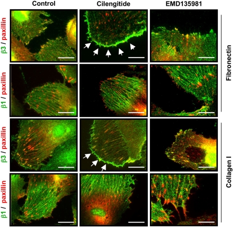
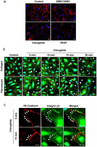
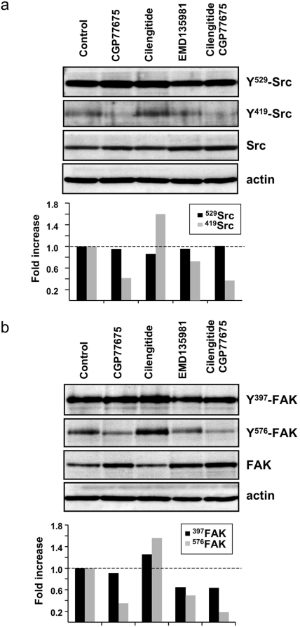
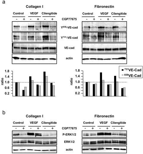
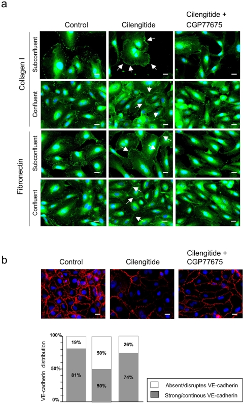
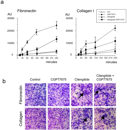
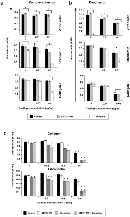
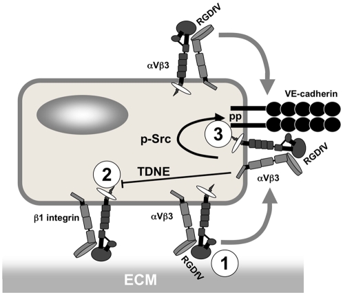
Cilengitide is a high-affinity cyclic pentapeptdic alphaV integrin antagonist previously reported to suppress angiogenesis by inducing anoikis of endothelial cells adhering through alphaVbeta3/alphaVbeta5 integrins. Angiogenic endothelial cells express multiple integrins, in particular those of the beta1 family, and little is known on the effect of cilengitide on endothelial cells expressing alphaVbeta3 but adhering through beta1 integrins. Through morphological, biochemical, pharmacological and functional approaches we investigated the effect of cilengitide on alphaVbeta3-expressing human umbilical vein endothelial cells (HUVEC) cultured on the beta1 ligands fibronectin and collagen I. We show that cilengitide activated cell surface alphaVbeta3, stimulated phosphorylation of FAK (Y(397) and Y(576/577)), Src (S(418)) and VE-cadherin (Y(658) and Y(731)), redistributed alphaVbeta3 at the cell periphery, caused disappearance of VE-cadherin from cellular junctions, increased the permeability of HUVEC monolayers and detached HUVEC adhering on low-density beta1 integrin ligands. Pharmacological inhibition of Src kinase activity fully prevented cilengitide-induced phosphorylation of Src, FAK and VE-cadherin, and redistribution of alphaVbeta3 and VE-cadherin and partially prevented increased permeability, but did not prevent HUVEC detachment from low-density matrices. Taken together, these observations reveal a previously unreported effect of cilengitide on endothelial cells namely its ability to elicit signaling events disrupting VE-cadherin localization at cellular contacts and to increase endothelial monolayer permeability. These effects are potentially relevant to the clinical use of cilengitide as anticancer agent.


Conflict of interest statement

Competing Interests: The authors have declared that no competing interests exist.

Figures

Figure 1
Figure 1. Cilengitide causes loss of αVβ3 from focal adhesions and promotes appearance of αVβ3 patches at the cell edge.
HUVEC were plated on coverslips coated with fibronectin or collagen I and were treated with 10 µM of cilengitide for 20 minutes. The localization of the αVβ3 or β1 integrin (green) and paxillin (red) were monitored by immunofluorescence staining. In HUVEC plated on fibronectin αVβ3 was present at focal adhesions, while β1 was present at fibrillar adhesions. Cilengitide, but not EMD 135981, caused loss of αVβ3 from focal adhesions and appearance of αVβ3-positive thin patches at the cell edge (arrows). β1 localization was not altered by cilengitide. A similar effect on αVβ3 (arrows) was observed on cells plated on collagen I, with the difference that focal adhesions were less abundant on this matrix. Optical magnification: 400×; Bar: 10 µm. (n = 5).
Figure 2
Figure 2. Cilengitide induces VE-cadherin loss form cellular junctions.
(a) Confluent HUVEC plated on fibronectin, were exposed to cilengitide or EMD135981 (10 µM each) or VEGF (100 ng/ml) for 20 minutes and stained for VE-cadherin. Cilengitide and VEGF treatments disrupted VE-cadherin localization at cellular junctions, while EMD135981 showed no effect (n = 3). Optical magnification: 400×; Bar: 10 µm. (b) Confluent HUVEC plated on fibronectin or collagen I were exposed to cilengitide (10 µM each) for the indicated time and double stained for VE-cadherin and β3 integrin. Cilengitide disrupted VE-cadherin staining and promoted appearance of β3 at VE-cadherin-depleted cell-cell borders (arrows). Paralleling loss of VE-cadherin from cell-cell junctions, ‘gaps’ appeared in the monolayer (asterisks). (n = 4). Optical magnification: 400×; Bar: 10 µm. (c) Higher magnification (2× zooming in) of HUVEC cultures of the experiment shown in panel b to demonstrate rare co-localization of VE-cadherin and β3 integrin at cellular junctions upon cilengitide stimulation (arrowheads). (n = 4). Bars: 10 µm.
Figure 3
Figure 3. Cilengitide activates αVβ3 on HUVEC.
(a) HUVEC in suspension were exposed to 10 µM of cilengitide or EMD135981 for 10 minutes, stained by immunofluorescence for β3 LIBS and total αVβ3 expression (with LIBS-1 and LM609 mAbs, respectively) and analyzed by flow cytometry. Cilengitide, but not EMD135981, induced LIBS expression (left histograms, thick lines), without affecting total αVβ3 expression (right histograms, thick lines). Dotted lines: cellular fluorescence in the absence of primary antibody. (n = 3). (b) Fibronectin-adherent HUVEC were exposed to 10 µM cilengitide, EMD135981, or 1 mM MnCl2 for 10 minutes, stained for β3 LIBS and total αVβ3 expression (with CRC54 or LM609 mAbs, respectively) and analyzed by immunofluorescence microscopy. Total αVβ3 and β3 LIBS were present at focal adhesions in unstimulated HUVEC and at tiny patches at the cell edge in cilengitide-exposed HUVEC, thus confirming that αVβ3-positive patches contain active αVβ3. MnCl2 stimulated recruitment and activation of αVβ3 at focal adhesions. (n = 2). Optical magnification: 400×; Bars: 10 µm.
Figure 4
Figure 4. Cilengitide induces Src and FAK phosphorylation.
(a) Western blotting analysis of Src phosphorylation at Y529 and Y419 and total Src in HUVEC grown on fibronectin and exposed for 10 minutes to EMD135981, cilengitide (10 µM each) and CGP77675 (2.5 µM) as indicated. Cilengitide increased Src phosphorylation at Y419 but did not alter Y529 phosphorylation. CGP77675 prevented Y419 phosphorylation. (b) Western blotting analysis of the same cells as in panel a, but for phosphorylation of FAK at Y397 and Y576 and total FAK. Cilengitide increased FAK phosphorylation at both tyrosine residues and this was inhibited by CGP77675. EMD135981 had no effect on Src or FAK phosphorylation. Actin was detected to demonstrate equal loading of the lanes. The bar graph represents the relative level of phospho Src/FAK over total Src/FAK as determined by band density analysis. (n = 3).
Figure 5
Figure 5. Cilengitide induces Src-dependent phosphorylation of VE-cadherin cytoplasmic domain.
(a) Western blotting analysis of VE-cadherin phosphorylation at tyrosine residues Y658 and Y731 and total VE-cadherin in HUVEC grown on fibronectin or collagen I and stimulated for 10 minutes with cilengitide (10 µM) or VEGF (100 ng/ml) in the presence or absence of CGP77675. Cilengitide treatment increased VE-cadherin phosphorylation at Y658 and Y731, while VEGF enhanced Y658 phosphorylation only. The bar graph represents the relative level of phospho VE-cadherin over total VE-cadherin as determined by band density analysis. (b) Western blotting analysis of phospho and total ERK 1/2 of the same cultures as in panel a. VEGF activated ERK 1/2, while cilengitide did not. Actin was detected to demonstrate equal loading of the lanes. (n = 3).
Figure 6
Figure 6. Src inhibition prevents cilengitide-induced relocalization of αVβ3 at the cell edge and disappearance of VE-cadherin from cellular junctions.
(a) Subconfluent and confluent HUVEC cultured on fibronectin or collagen I were exposed for 20 minutes to cilengitide (10 µM) in the presence of absence of CGP77675 (2.5 µM) and stained for αVβ3. Cilengitide-induced recruitment of αVβ3 to the cell edge (arrows) and this was prevented by CGP77675. (b) Confluent HUVEC cultured on fibronectin were treated for 20 minutes with cilengitide in the presence or absence of CGP77675. CGP77675 prevented cilengitide-induced VE-cadherin loss from cell-cell contacts. The bar graph gives the quantification of VE-cadherin staining at cell borders. The white and gray segments of the bars represent absent/disrupted vs. strong/continuous VE-cadherin staining, respectively (see material and methods for details). (n = 3). Optical magnification: 400×; Bars: 10 µM.
Figure 7
Figure 7. Cilengitide augments the permeability of HUVEC monolayers.
(a). HUVEC were grown on fibronectin- or collagen I-coated PET filter inserts for 20 hours to ensure confluence and treated with cilengitide (10 µM), CGP77675 (2.5 µM) or a combination thereof. Permeability was measured using the tracer molecule FITC-dextran. Cilengitide increased HUVEC monolayer permeability on both matrices and CGP77675 only partially prevented this increase. Results represent the increase in permeability of treated cultures relative to untreated controls at t = 0 and is given in arbitrary fluorescence units (AU). (b) Crystal violet staining of control and treated filters at the end of the assay (240 minutes) revealed that cilengitide did not cause extensive detachment of HUVEC but induced the appearance of retraced, dendritic-like cells (arrows). (Triplicate filters/condition, n = 3).
Figure 8
Figure 8. Cilengitide interferes with HUVEC adhesion on low-density β1 integrin substrates.
(a) HUVEC short-term adhesion assays performed on vitronectin (αVβ3 ligand), fibronectin (α5β1>αVβ3 ligand) and collagen I (α1β1/α2β1 ligand) coated at the indicated concentrations, in medium only (black bars) or in the presence of EMD135981 (gray bars) or cilengitide (white bars). On vitronectin, cilengitide inhibited adhesion at all coating concentrations while on fibronectin and collagen I it blocked adhesion only at low coating concentrations. (n = 5). (b) HUVEC detachment assays. HUVEC were cultured for 18 hours on vitronectin, fibronectin or collagen I coated at the indicated concentrations, to allow for full attachment, before exposure for 4 hours to medium only (black bars), EMD135981 (gray bars) or cilengitide (white bars). Cilengitide detached HUVEC cultured on vitronectin at all coating concentrations, while it induced HUVEC detachment from fibronectin and collagen only at low coating concentrations (Triplicate wells/condition, n = 3). (c) HUVEC short-term adhesion assays performed on fibronectin and collagen I coated at the indicated concentrations, in medium only (black bars), in the presence of CGP77675 (white bars), cilengitide (gray bars), cilengitide+CGP77675 (hatched bars). Src inhibition did not prevent cilengitide-induced inhibition of cell adhesion on low matrix concentrations. (Triplicate wells/condition, n = 2). Attached cells were quantified by Crystal Violet staining and OD determination at 540 nm wavelength. Asterisks indicate statistical significant differences of the values relative to untreated controls (p<0.05).
Figure 9
Figure 9. Proposed model of cilengitide effects on endothelial cells.
Cilengitide acts on αVβ3-expressing endothelial cells in three ways: 1, it suppresses αVβ3-dependent adhesion by directly inhibiting αVβ3-ligand-binding function; 2, it interferes with β1 integrin-mediated cell adhesion through a transdominant negative effect induced by activated αVβ3; 3, it stimulates phosphorylation of VE-cadherin cytoplasmic domain and disrupts VE-cadherin localization at cell-cell contacts through activation of αVβ3 and Src-dependent signaling. Abbreviations: ECM, extracellular matrix; RGDfV, cilengitide; TDNE, transdominant negative effect; pp, phosphorylation.

References

    1. Hynes RO. Cell-matrix adhesion in vascular development. J Thromb Haemost. 2007;5(Suppl 1):32–40. - PubMed
    1. Hynes RO. Integrins: bidirectional, allosteric signaling machines. Cell. 2002;110:673–687. - PubMed
    1. Alghisi GC, Rüegg C. Vascular Integrins in Tumor Angiogenesis: Mediators and Therapeutic Targets. Endothelium. 2006;13:113–135. - PubMed
    1. Kumar CC. Integrin alphavbeta3 as a Therapeutic Target for Blocking Tumor-Induced Angiogenesis. Current Drug Targets. 2003;4:123–131. - PubMed
    1. Smith J. Cilengitide Merck. Curr Opin Investig Drugs. 2003;4:741–745. - PubMed

Publication types

MeSH terms