. 2009;4(2):e4440.
doi: 10.1371/journal.pone.0004440. Epub 2009 Feb 12.

Inoculation of scrapie with the self-assembling RADA-peptide disrupts prion accumulation and extends hamster survival

Affiliations

Inoculation of scrapie with the self-assembling RADA-peptide disrupts prion accumulation and extends hamster survival

Robert Hnasko et al. PLoS One. 2009.

Abstract

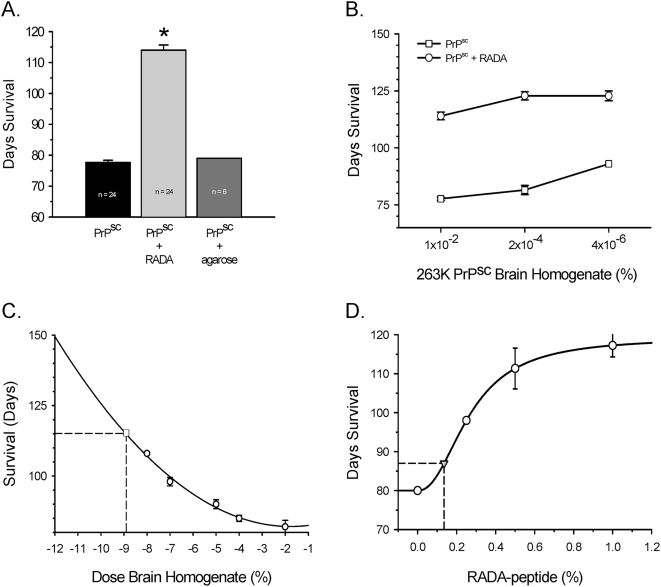
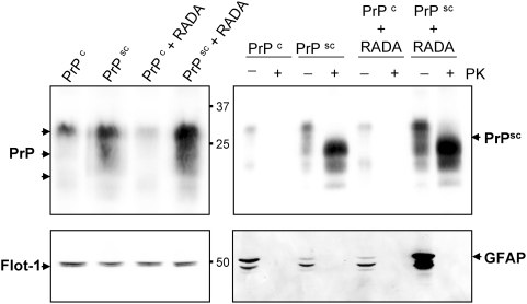
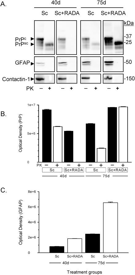
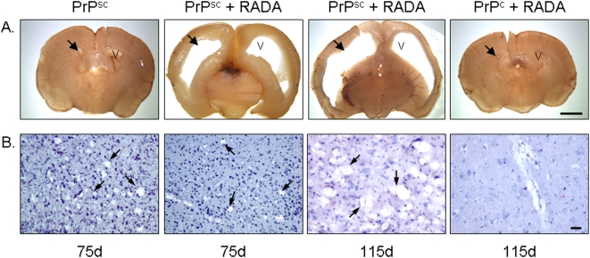
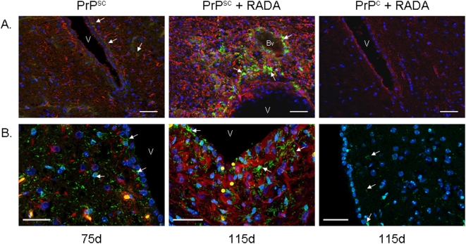
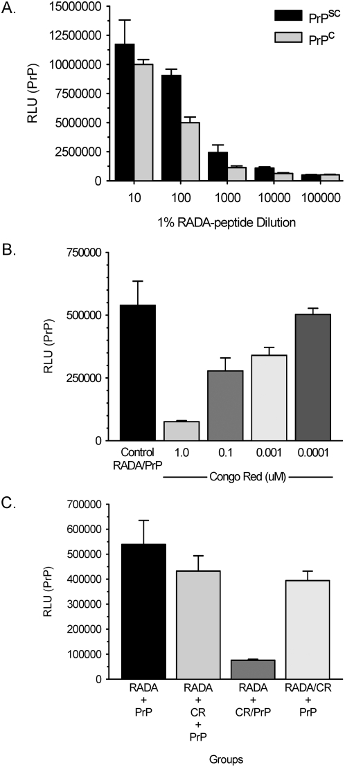
Intracerebral inoculation of 263K Scrapie brain homogenate (PrPsc) with a self-assembling RADA-peptide (RADA) significantly delayed disease onset and increased hamster survival. Time of survival was dependent on the dose of RADA and pre-incubation with PrPsc prior to inoculation. RADA treatment resulted in the absence of detectable PrPsc at 40 d followed by an increased rate of PrPsc accumulation at 75 d up to sacrifice. In all PrPsc inoculated animals, clinical symptoms were observed approximately 10 d prior to sacrifice and brains showed spongiform degeneration with Congo red positive plaques. A time-dependent increase in reactive gliosis was observed in both groups with more GFAP detected in RADA treated animals at all time points. The PrP protein showed dose-dependent binding to RADA and this binding was competitively inhibited by Congo Red. We conclude that RADA disrupts the efficacy of prion transmission by altering the rate of PrPsc accumulation. This is the first demonstration that a self-assembling biomolecular peptide can interact with PrPsc, disrupt the course of Scrapie disease process, and extend survival.


Conflict of interest statement

Competing Interests: The authors have declared that no competing interests exist.

Figures

Figure 1
Figure 1. RADA promotes increased survival of hamsters inoculated with 263K Scrapie.
Fig. 1A compares hamster survival in days (mean±SEM) following intracerebral inoculation with 1% Scrapie alone (PrPsc), Scrapie pre-incubated with 0.9% w/v RADA (PrPsc+RADA) or Scrapie agarose plugs (PrPsc+agarose). Fig. 1B compares hamster survival in days (mean±SEM) with increased dilution of Scrapie brain homogenate alone (open squares) to equivalent doses of Scrapie pre-incubated with 0.9% RADA (open circles). Fig. 1C depicts a hamster survival curve in days (N = 6; mean±SEM) with increasing dilution of Scrapie inoculant (open circles). Plotted on the curve (open square) is the mean survival in days of hamsters inoculated with 10−2 Scrapie combined with 0.9% RADA depicting equivalent titer of Scrapie inoculum. Fig. 1D shows a dose-dependent increase in hamster survival in days (mean±SEM) with increasing concentration of RADA (open circles) inoculated with 1% Scrapie. A synthetic 16-mer RADA-peptide was hydrated combined with 1% Scrapie and inoculated. Survival in days was plotted (grey triangle) and used to estimate RADA concentration.
Figure 2
Figure 2. Prion and GFAP proteins are increased in brain homogenates from hamsters inoculated with Scrapie+RADA.
Total PrP protein was detected by Western blot in hamster brain homogenates (20 µg/lane) from normal (PrPc; 75 d), Scrapie infected (PrPsc; 75 d), normal combined with RADA (PrPc+RADA; 115 d), and Scrapie combined with RADA (PrPsc+RADA; 115 d). A prominent PrP band was detected at ∼30 kDa and two lower molecular weight bands at ∼22 kDa and 19 kDa (top left panel). Flotillin-1 (Flot-1) was used as a loading control and a doublet was detected at ∼45 kDa (bottom left panel). Digestion of brain homogenates with proteinase-K (+PK) demonstrated the presence of prion protein in the PrPsc and PrPsc+RADA samples, but not in PrPc or PrPc+RADA. A detectable MW shift was observed for PrPsc in all three bands with a shift from 30 kDa to 25 kDa occurring for the predominant band (top right panel). A notable increase in PK-resistant prion was detected in the PrPsc+RADA compared to the PrPsc alone. Detection of GFAP was used to verify the complete PK-digestion of samples (bottom right panel). Three detectable GFAP bands were resolved (∼50, 45 and 40 kDa) in the non-PK treated samples with increased detection of GFAP in PrPsc+RADA brain.
Figure 3
Figure 3. Time-dependent increase in GFAP and PrP protein in hamsters inoculated with combined Scrapie+RADA.
Fig. 3A shows a Western blot from hamster brain homogenates inoculated with 1% Scrapie alone (Sc) or combined with 0.9% RADA (Sc+RADA) at 40 d and 75 d treated with (+) or without (−) proteinase-K (PK). The top panels show PrPc and PrPsc detection at each time point (20 µg). Middle panels show detection of GFAP protein and bottom panel contactin-1. Fig. 3B shows quantification of total PrP (−PK) and prion (+PK) from Western blots at each time-point. Fig. 3C show quantification of ∼55 kDa GFAP band (−PK) from Western blots from each time-point.
Figure 4
Figure 4. Combined inoculation of Scrapie+RADA results in ventricular enlargement and delayed spongiform degeneration.
Brain sections were obtained from hamsters inoculated with 1% Scrapie brain homogenate alone (PrPsc) after 75 d, Scrapie combined with 0.9% RADA (PrPsc+RADA) after 75 d and 115 d and normal brain combined with RADA (PrPc+RADA) after 115 d. Fig. 4A shows gross morphology from coronal sections of whole hamster brains. No ventricular enlargement (black arrows) was observed in animals that received inoculation of PrPsc alone (top left panel) or PrPc+RADA (top right panel), whereas brains from hamsters inoculated with PrPsc+RADA had massive enlargement of lateral ventricles at both 75 d and 115 d (top middle panels). V = ventricle. Bar = 2 mm. Fig. 4B are micrographs from brain sections stained with Hematoxylin showing spongiform degeneration present in hamsters cortex following inoculation with PrPsc alone after 75 d (bottom left panel; arrows) and PrPsc+RADA after 75 d and 115 d (middle panels; arrows). No spongiform degeneration was detected in animals inoculated with PrPc+RADA after 115 d (bottom right panel). Bar = 50 µm.
Figure 5
Figure 5. Scrapie induced reactive gliosis and PrP aggregation is increased by RADA.
Fig. 5A shows dual labeled immunofluorescent GFAP (red) and PrP (green) hamster brain micrographs. Limited staining of GFAP and no detectable PrP was observed in PrPc+RADA treated brain after 115 d (top right panel). PrPsc treatment alone results in an increased detection of GFAP-positive astrocytes (red) and weak PrP protein (green, white arrows) after 75 d (middle panel). Combined PrPsc+RADA treatment (right panel) results in abundant and strong detection of both GFAP-positive astrocytes and large PrP deposits (green, arrows). V = ventricle. Bv = blood vessel. DAPI nuclei (blue). Bars = 50 µm. Fig. 5B shows micrographs of hamster brain sections labeled with GFAP (red) and IBA1 (green). Increased IBA1-positive immunoreactive microglia are detected in brain following inoculation of PrPsc alone after 75 d (left panel; white arrows) and PrPsc+RADA after 115 d (middle panel; white arrows) as compared to PrPc+RADA after 115 d (right panel; white arrows). IBA1 and GFAP immunoreactivity do not co-localize in dual labeled sections from PrPsc and PrPsc+RADA brain. V = ventricle. DAPI nuclei (blue). Bars = 50 µm.
Figure 6
Figure 6. PrP binds RADA-peptide and binding can be inhibited with Congo red.
Fig. 6A shows chemiluminescent detection of PrP binding to RADA dilutions from Scrapie (PrPsc) and normal (PrPc) brain homogenates. Fig. 6B shows quantification of PrP binding to RADA (Control RADA/PrP) following pre-incubation with dilutions of Congo Red. Fig. 6C compares the binding of PrP to RADA based on the order of reagent addition. Quantification of PrP binding to RADA: RADA followed by PrP (RADA+PrP), RADA followed by 1 µM Congo red (CR) then PrP (RADA+CR+PrP), RADA followed by pre-incubated PrP with 1 µM CR (RADA+CR/PrP), RADA pre-incubated with 1 µM CR followed by PrP (RADA/CR+PrP). RLU = relative light units.

Similar articles

Cited by

References

    1. Prusiner SB. Prions. Proc Natl Acad Sci U S A. 1998;95:13363–13383. - PMC - PubMed
    1. Bolton DC, McKinley MP, Prusiner SB. Identification of a protein that purifies with the scrapie prion. Science. 1982;218:1309–1311. - PubMed
    1. Prusiner SB, Groth D, Serban A, Koehler R, Foster D, et al. Ablation of the prion protein (PrP) gene in mice prevents scrapie and facilitates production of anti-PrP antibodies. Proc Natl Acad Sci U S A. 1993;90:10608–10612. - PMC - PubMed
    1. DeArmond SJ, Yang SL, Cayetano-Canlas J, Groth D, Prusiner SB. The neuropathological phenotype in transgenic mice expressing different prion protein constructs. Philos Trans R Soc Lond B Biol Sci. 1994;343:415–423. - PubMed
    1. Mallucci G, Dickinson A, Linehan J, Klohn PC, Brandner S, et al. Depleting neuronal PrP in prion infection prevents disease and reverses spongiosis. Science. 2003;302:871–874. - PubMed

Publication types