

Multiple hereditary osteochondromatosis: a case report
- PMID: 19212564
- PMCID: PMC2638239
Multiple hereditary osteochondromatosis: a case report
Abstract
Objectives: Common carious lesions owing to vomiting are not widespread in children. In this case, we aimed to report an 11-years-old male patient with common carious lesions due to repeated vomitings, chewing and eating difficulty and retarded growth with Multiple Hereditary Osteochondromatosis (MHO).
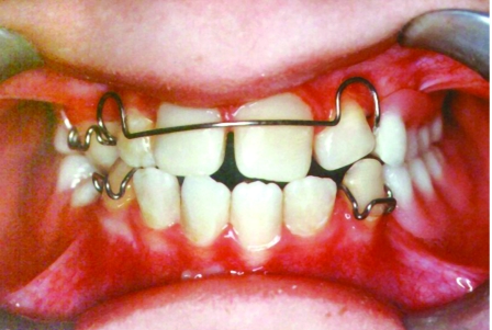
Case report: An 11-years-old boy was referred to Department of Pediatric Dentistry in Faculty of Dentistry because of eating difficulty owing to common carious lesions. It was seen that the patient growth was generally retarded in extra-oral examination. Some exostoses were also present on the extremities. It was learned that he was previously diagnosed as MHO in Faculty of Medicine. Nausea and vomiting have been commonly occurring after taking of Didronat. Chewing and eating difficulty and inadequate nutrition were present because of bad oral hygiene, carious lesions and remained roots. Growth was negatively affected by malnutrition and MHO.
Results: Diet recommendations were given and oral hygiene behaviors were rearranged. Preventive, surgical, restorative and prosthetical dental applications were applied for dental treatments.
Keywords: Bisphosphonates; Bone tumor; Chondrosarcoma; Hereditary multiple osteochondromatosis; Multiple hereditary exostoses.
Figures


Similar articles
-
Accuracy of pediatric primary care providers' screening and referral for early childhood caries.Pediatrics. 2002 May;109(5):E82-2. doi: 10.1542/peds.109.5.e82. Pediatrics. 2002. PMID: 11986488
-
Dedifferentiated chondrosarcoma in patients with multiple osteochondromatosis: report of a case and review of the literature.Skeletal Radiol. 1997 Jun;26(6):370-4. doi: 10.1007/s002560050250. Skeletal Radiol. 1997. PMID: 9229421 Review.
-
An unusual complication in a 9-year-old patient with hereditary multiple osteochondromatosis.PM R. 2013 Apr;5(4):348-50. doi: 10.1016/j.pmrj.2013.01.014. PM R. 2013. PMID: 23622788
-
Determination of bone age in children with cartilaginous dysplasia (multiple hereditary osteochondromatosis and Ollier's enchondromatosis).J Pediatr Orthop. 2004 Jan-Feb;24(1):102-8. doi: 10.1097/00004694-200401000-00020. J Pediatr Orthop. 2004. PMID: 14676544
-
Acetabular dysplasia associated with hereditary multiple exostoses. A case report.J Bone Joint Surg Br. 2000 May;82(4):555-7. doi: 10.1302/0301-620x.82b4.9894. J Bone Joint Surg Br. 2000. PMID: 10855881 Review.
Cited by
-
Multiple hereditary osteochondromatosis with spinal cord compression: case report.Childs Nerv Syst. 2018 Mar;34(3):565-569. doi: 10.1007/s00381-017-3645-1. Epub 2017 Nov 11. Childs Nerv Syst. 2018. PMID: 29129004
-
HSPG-deficient zebrafish uncovers dental aspect of multiple osteochondromas.PLoS One. 2012;7(1):e29734. doi: 10.1371/journal.pone.0029734. Epub 2012 Jan 11. PLoS One. 2012. PMID: 22253766 Free PMC article.
References
-
- Noonan KJ, Levenda A, Snead J, Feinberg JR, MIH A. Evaluation of the forearm in untreated adult subjects with multiple hereditary osteochondromatosis. The Journal of Bone & Joint Surgery. 2002;84-A:397–403. - PubMed
-
- Peterson HA. Multiple Hereditary Osteochondromata. Clin Orthop. 1989;239:222–230. - PubMed
-
- Porter DE, Emerton ME, Villanueva-Lopez F, Simpson AH. Clinical and radiographic analysis of osteochondromas and growth disturbance in hereditary multiple exostoses. J Pediatr Orthop. 2000;20:246–250. - PubMed
-
- Vanhoenacker FM, Van Hul W, Wuyts W, Willems PJ, De Schepper AM. Hereditary multiple exostoses: from genetics to clinical syndrome and complications. Eur J Radiol. 2001;40:208–217. - PubMed
-
- Faiyaz-Ul-Haque M, Ahmad W, Zaidi SHE, Hussain S, Haque S, Ahmad M, et al. Novel mutations in the EXT1 gene in two consanguineous families affected with multiple hereditary exostoses (familial osteochondromatosis) Clin Genet. 2004;66:144–151. - PubMed
Publication types
LinkOut - more resources
Full Text Sources
Research Materials