. 2009 Mar;23(3):389-97.
doi: 10.3892/ijmm_00000143.

Mutational analysis of the cleavage of the cancer-associated laminin receptor by stromelysin-3 reveals the contribution of flanking sequences to site recognition and cleavage efficiency

Affiliations

Mutational analysis of the cleavage of the cancer-associated laminin receptor by stromelysin-3 reveals the contribution of flanking sequences to site recognition and cleavage efficiency

Maria Fiorentino et al. Int J Mol Med. 2009 Mar.

Abstract

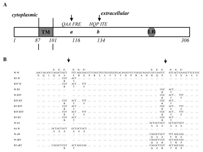
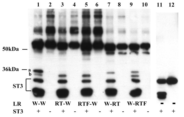
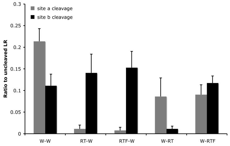
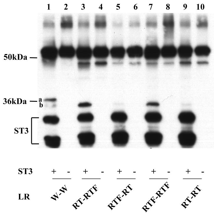
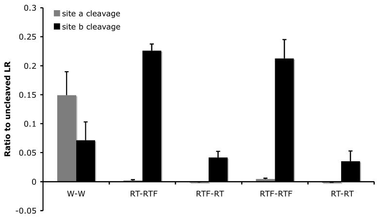
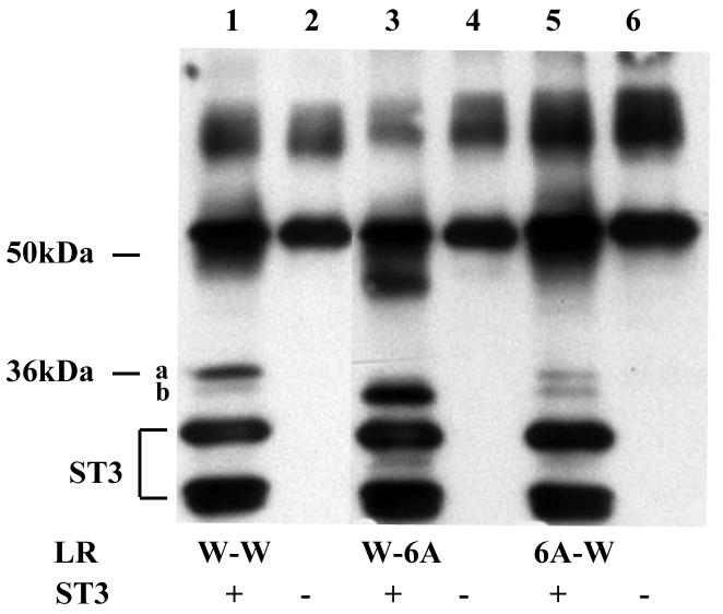
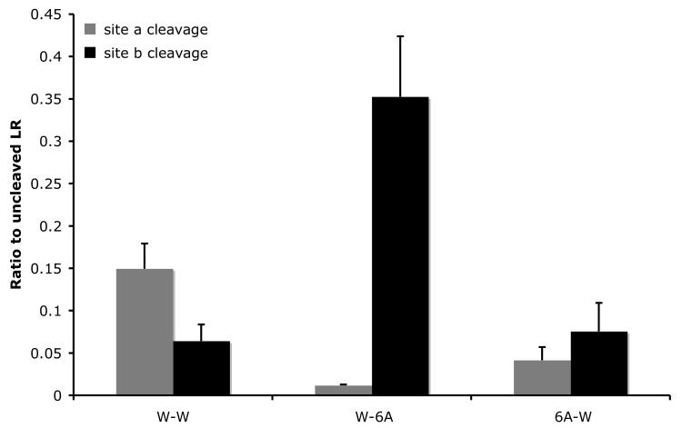
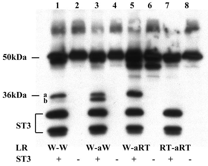
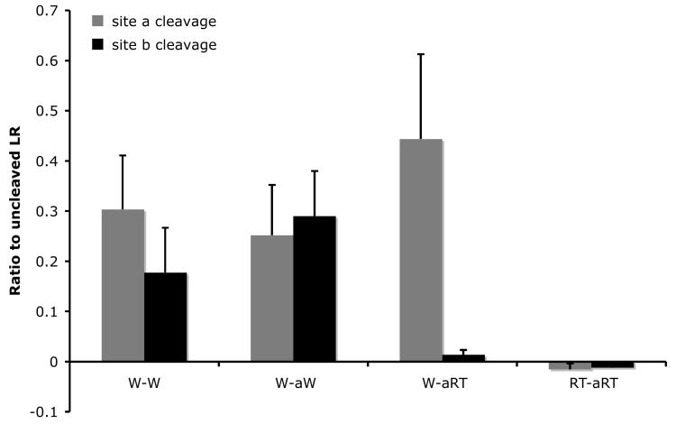
The matrix metalloproteinase stromelysin-3 (ST3) has long been implicated to play an important role in cell fate determination during normal and pathological processes. Using the thyroid hormone-dependent Xenopus laevis metamorphosis as a model, we have previously shown that ST3 is required for apoptosis during intestinal remodeling and that laminin receptor (LR) is an in vivo substrate of ST3 during this process. ST3 cleaves LR at two distinct sites that are conserved in mammalian LR. Human ST3 and LR are both associated with tumor development and cancer progression and human LR can also be cleaved by ST3, implicating a role of LR cleavage by ST3 in human cancers. Here, we carried out a series of mutational analyses on the two cleavage sites in LR. Our findings revealed that in addition to primary sequence at the cleavage site (positions P3-P3', with the cleavage occurring between P1-P1'), flanking sequences/conformation also influenced the cleavage of LR by ST3. Furthermore, alanine substitution studies led to a surprising finding that surrounding sequence and/or conformation dictated the site of cleavage in LR by ST3. These results thus have important implications in our understanding of substrate recognition and cleavage by ST3 and argue for the importance of studying ST3 cleavage in the context of full-length substrates. Furthermore, the LR cleavage mutants generated here will also be valuable tools for future studies on the role of LR cleavage by ST3 in vertebrate development and cancer progression.


Figures

Fig. 1
Fig. 1
A. Schematic diagram showing ST3 cleavage sites between the transmembrane domain (TM) and the laminin binding sequence (LB) of LR (27). ST3 cleaves LR between A115 and F116 (site a), and P133 and I134 (site b) and these two cleavage sites by ST3 are indicated by two arrows (Based on (27)). B. LR mutants used in this study.
Fig. 2
Fig. 2
Differential effects of amino acid substitutions on the cleavage of the two sites in LR by ST3. A. RT and RTF mutations at site a essentially abolished ST3 cleavage at this site (compare lanes 3 and 5, respectively, to lane 1). While RT mutation at site b also inhibited ST3 cleavage at this site (lane 7), an additional substitution of E to F at the P3′ position restored the ability of ST3 to cleave at this site (compare lane 1 to lane 9). His-tagged wild type LR and indicated mutants were synthesized by in vitro translation, purified, and digested with purified Xenopus ST3 catalytic domain. The samples were mixed with SDS sample buffer and subjected to Western blotting with anti-Xenopus LR antibody. Note that the anti-LR antibody was made against His-tagged LR and also recognizes His-tagged ST3. During the incubation, ST3 auto degraded to produce the lower molecular weight bands that were present both in the presence (lanes 1, 3, 5, 7, 9) or absence of LR (11) but not in the ST3 sample without incubation (12) (lanes 11 and 12 were from a separate gel). The figure is a representative of 2 independent experiments with similar results. B. Quantification of the data shown in Fig. A. Shown here is the average from 2 independent experiments.
Fig. 2
Fig. 2
Differential effects of amino acid substitutions on the cleavage of the two sites in LR by ST3. A. RT and RTF mutations at site a essentially abolished ST3 cleavage at this site (compare lanes 3 and 5, respectively, to lane 1). While RT mutation at site b also inhibited ST3 cleavage at this site (lane 7), an additional substitution of E to F at the P3′ position restored the ability of ST3 to cleave at this site (compare lane 1 to lane 9). His-tagged wild type LR and indicated mutants were synthesized by in vitro translation, purified, and digested with purified Xenopus ST3 catalytic domain. The samples were mixed with SDS sample buffer and subjected to Western blotting with anti-Xenopus LR antibody. Note that the anti-LR antibody was made against His-tagged LR and also recognizes His-tagged ST3. During the incubation, ST3 auto degraded to produce the lower molecular weight bands that were present both in the presence (lanes 1, 3, 5, 7, 9) or absence of LR (11) but not in the ST3 sample without incubation (12) (lanes 11 and 12 were from a separate gel). The figure is a representative of 2 independent experiments with similar results. B. Quantification of the data shown in Fig. A. Shown here is the average from 2 independent experiments.
Fig. 3
Fig. 3
Mutations at site a enhance the cleavage of mutant site b by ST3. A. By inhibiting ST3 cleavage at site a with RT or RTF mutations, the cleavage at site b with RT or RTF mutations was enhanced compared to the wild-type LR (compare lane 1 to lanes 3 and 7) and to the single mutants (see Fig. 2). His-tagged wild type LR and indicated mutants were synthesized by in vitro translation, purified, and digested with purified Xenopus ST3 catalytic domain. The samples were subjected to Western blotting with anti-Xenopus LR antibody. The figure is a representative of 2 independent experiments. B. Quantification of the data shown in A. Shown here is the average from 2 independent experiments.
Fig. 3
Fig. 3
Mutations at site a enhance the cleavage of mutant site b by ST3. A. By inhibiting ST3 cleavage at site a with RT or RTF mutations, the cleavage at site b with RT or RTF mutations was enhanced compared to the wild-type LR (compare lane 1 to lanes 3 and 7) and to the single mutants (see Fig. 2). His-tagged wild type LR and indicated mutants were synthesized by in vitro translation, purified, and digested with purified Xenopus ST3 catalytic domain. The samples were subjected to Western blotting with anti-Xenopus LR antibody. The figure is a representative of 2 independent experiments. B. Quantification of the data shown in A. Shown here is the average from 2 independent experiments.
Fig. 4
Fig. 4
Alanine substitution from P3-P3′ inhibits cleavage at site a but enhances that at site b. A. Six alanine (6A) were introduced to replace the 3 amino acids on either side of each cleavage site. His-tagged wild type LR and indicated mutants were synthesized by in vitro translation, purified, and digested with purified Xenopus ST3 catalytic domain. The samples were subjected to Western blotting with anti-Xenopus LR antibody. Note that all 6 lanes were run on the same gel but two lanes for a construct not relevant to this figure were removed. The figure is a representative of 2 independent experiments with similar results. B. Quantification of the data shown in A. Shown here is the average from 2 independent experiments.
Fig. 4
Fig. 4
Alanine substitution from P3-P3′ inhibits cleavage at site a but enhances that at site b. A. Six alanine (6A) were introduced to replace the 3 amino acids on either side of each cleavage site. His-tagged wild type LR and indicated mutants were synthesized by in vitro translation, purified, and digested with purified Xenopus ST3 catalytic domain. The samples were subjected to Western blotting with anti-Xenopus LR antibody. Note that all 6 lanes were run on the same gel but two lanes for a construct not relevant to this figure were removed. The figure is a representative of 2 independent experiments with similar results. B. Quantification of the data shown in A. Shown here is the average from 2 independent experiments.
Fig. 5
Fig. 5
Substitution of the six amino acids at the cleavage site b with the corresponding ones at site a creates a cleavage site with site a characteristics. A. The 3 amino acids at either side of site b were replaced with the corresponding amino acids at site a to generate W-aW. RT mutations were then introduced into either one or both of the cleavage sites in W-aW to create the other mutants. His-tagged wild type LR and indicated mutants were synthesized by in vitro translation, purified, and digested with purified Xenopus ST3 catalytic domain. The samples were subjected to Western blotting with anti-Xenopus LR antibody. Note that RT mutation in the site b with the site a sequence completely blocked the cleavage by ST3 and RT mutation in site a failed to enhance the cleavage at this mutant site b (lane 7), in contrast to the corresponding mutants made from the wild type LR (Fig. 3, lane 9). The figure is a representative of 2 independent experiments with similar results. B. Quantification of the data shown in A. Shown here is the average from 2 independent experiments.
Fig. 5
Fig. 5
Substitution of the six amino acids at the cleavage site b with the corresponding ones at site a creates a cleavage site with site a characteristics. A. The 3 amino acids at either side of site b were replaced with the corresponding amino acids at site a to generate W-aW. RT mutations were then introduced into either one or both of the cleavage sites in W-aW to create the other mutants. His-tagged wild type LR and indicated mutants were synthesized by in vitro translation, purified, and digested with purified Xenopus ST3 catalytic domain. The samples were subjected to Western blotting with anti-Xenopus LR antibody. Note that RT mutation in the site b with the site a sequence completely blocked the cleavage by ST3 and RT mutation in site a failed to enhance the cleavage at this mutant site b (lane 7), in contrast to the corresponding mutants made from the wild type LR (Fig. 3, lane 9). The figure is a representative of 2 independent experiments with similar results. B. Quantification of the data shown in A. Shown here is the average from 2 independent experiments.

Similar articles

Cited by

References

    1. Barrett JA, Rawloings ND, Woessner JF. Handbook of proteolytic enzymes. Academic Press; NY: 1998.
    1. Alexander CM, Werb Z. Extracellular matrix degradation. In: Hay ED, editor. Cell Biology of Extracellular Matrix. 2nd ed. Plenum Press; New York: 1991. pp. 255–302.
    1. Birkedal-Hansen H, Moore WGI, Bodden MK, et al. Matrix metalloproteinases: a review. Crit. Rev. in Oral Biol. and Med. 1993;4:197–250. - PubMed
    1. McCawley LJ, Matrisian LM. Matrix metalloproteinases: they're not just for matrix anymore! Current Opinion in Cell Biology. 2001;13:534–540. - PubMed
    1. Parks WC, Mecham RP. Matrix metalloproteinases. Academic Press; New York: 1998.

Publication types