. 2009 Apr 10;284(15):10276-84.
doi: 10.1074/jbc.M806907200. Epub 2009 Feb 12.

Location of the antidepressant binding site in the serotonin transporter: importance of Ser-438 in recognition of citalopram and tricyclic antidepressants

Affiliations

Location of the antidepressant binding site in the serotonin transporter: importance of Ser-438 in recognition of citalopram and tricyclic antidepressants

Jacob Andersen et al. J Biol Chem. .

Abstract

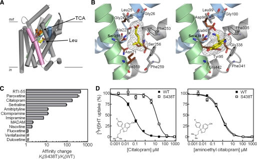
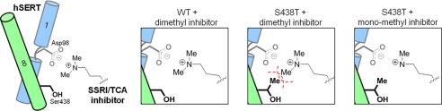
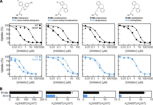
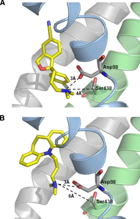
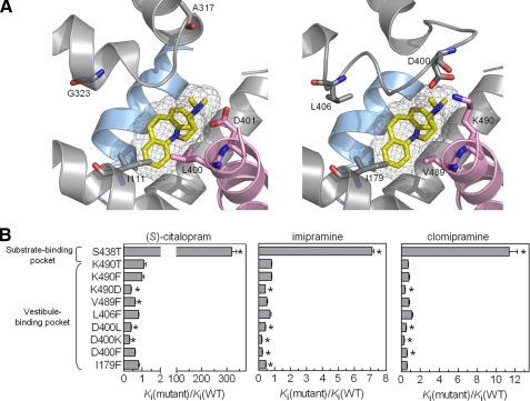
The serotonin transporter (SERT) regulates extracellular levels of serotonin (5-hydroxytryptamine, 5HT) in the brain by transporting 5HT into neurons and glial cells. The human SERT (hSERT) is the primary target for drugs used in the treatment of emotional disorders, including depression. hSERT belongs to the solute carrier 6 family that includes a bacterial leucine transporter (LeuT), for which a high resolution crystal structure has become available. LeuT has proved to be an excellent model for human transporters and has advanced the understanding of solute carrier 6 transporter structure-function relationships. However, the precise structural mechanism by which antidepressants inhibit hSERT and the location of their binding pockets are still elusive. We have identified a residue (Ser-438) located within the 5HT-binding pocket in hSERT to be a critical determinant for the potency of several antidepressants, including the selective serotonin reuptake inhibitor citalopram and the tricyclic antidepressants imipramine, clomipramine, and amitriptyline. A conservative mutation of Ser-438 to threonine (S438T) selectively increased the K(i) values for these antidepressants up to 175-fold. The effects of introducing a protein methyl group into the 5HT-binding pocket by S438T were absent or reduced for analogs of these antidepressants lacking a single methyl group. This suggests that these antidepressants interact directly with Ser-438 during binding to hSERT, implying an overlapping localization of substrate- and inhibitor-binding sites in hSERT suggesting that antidepressants function by a mechanism that involves direct occlusion of the 5HT-binding site.


Figures

FIGURE 1.
FIGURE 1.
Topology of SERT derived from LeuT. The substrate-binding pocket is formed by residues positioned in TM1, TM3, TM6, and TM8 (shown in gray). Residues elucidated as critical in recognition of SSRIs and TCAs are indicated as follows: Tyr-95 (29), Asp-98 (23), Ala-169 (31), Ile-172 (28), Met-180 (30), Ser-276 (40), Phe-513 (30), Ser-545 (41), and Phe-586 (42).
FIGURE 2.
FIGURE 2.
Mutation of Ser-438 in the hSERT substrate-binding pocket affects the inhibitory potency of antidepressants. A, structure of LeuT in complex with leucine and desipramine (PDB code 2QJU). TM1 (blue), TM8 (green), and TM10 (pink) are highlighted. TM6 and TM11 are removed for clarity. Leucine and desipramine are shown as CPK representations in yellow and orange, respectively. Arg-30 and Asp-404 (shown as sticks) form the extracellular gate that separates leucine from the bound desipramine. B, left, substrate-binding site in LeuT (PDB code 2A65). Right, homology model of the substrate-binding site in hSERT. Conserved residues are shown in gray, and divergent residues are shown in dark red. TM1 (blue), TM6 (gray), and TM8 (green) are shown in both panels. C, graphical summary of the fold change (mean ± S.E.; n = 6-8) in potency of inhibitors at hSERT S438T compared with hSERT WT. Ki values and statistics are shown in Table 1 and supplemental Table S1. # indicates that fold change is less than 1. D, inhibition of 5HT uptake at hSERT WT (▪) and S438T (□) by citalopram and aminoethyl citalopram. The concentration-response curve as shown is a composite of six independent experiments. Error bars are S.E. and are shown when larger than symbol size.
FIGURE 3.
FIGURE 3.
Proposed working model for the impact of S438T mutation. Schematic showing hSERT interaction with the aminopropyl chain of TCAs and SSRIs. Asp-98 on TM1 (blue) coordinates the aminopropyl group. Introduction of a methyl group at Ser-438 on TM8 (green) by the S438T mutation leads to steric clash (indicated by the red dashed line) with a methyl group on the aminopropyl chain. The steric clash is removed with monomethyl analogs.
FIGURE 4.
FIGURE 4.
Functional [3H]5HT uptake inhibition analysis at hSERT WT and hSERT S438T. A, upper, structure of inhibitors having one (R = H) or two (R = CH3) methyl groups at the aminopropyl chain. Lower, characterization of pairs of inhibitors having a single (blue squares, WT; blue triangles, S438T) or two (▪, WT; ▴, S438T) methyl groups at the aminopropyl chain. B, graphical summary of observed fold change in Ki between WT and S438T for each inhibitor pair (mean ± S.E.; n = 6-8). Ki values and statistics are shown in Table 1.
FIGURE 5.
FIGURE 5.
Induced fit docking of (S)-citalopram (A) and imipramine (B). The highest scoring binding modes of both compounds are shown. TM1 (blue), TM3 and TM8 (green) are shown in both panels. Distances between the amino groups of the inhibitors and the carboxylate oxygen atom of Asp-98 and the C-β atom of Ser-438 are shown.
FIGURE 6.
FIGURE 6.
Impact of vestibule mutations in hSERT on inhibitor potency. A, left, structure of the TCA-binding site in LeuT with imipramine bound (PDB code 2Q72). Right, overlay of homology model of hSERT with LeuT structure (shown in left panel) to show the equivalent site in hSERT. TM1 (blue) and TM3, TM6, EL4, and TM10 (pink) are shown. B, graphical summary of fold change (mean ± S.E.; n = 6-10) in potency of (S)-citalopram, imipramine, and clomipramine at hSERT mutants compared with WT hSERT. For comparison, the fold changes in potency for (S)-citalopram, imipramine, and clomipramine at S438T compared with WT hSERT are shown. Ki values and statistics are shown in supplemental Table S2. * indicates a significant change (p < 0.05) in Ki values.

References

    1. Wong, D. T., Perry, K. W., and Bymaster, F. P. (2005) Nat. Rev. Drug Discov. 4 764-774 - PubMed
    1. Yamashita, A., Singh, S. K., Kawate, T., Jin, Y., and Gouaux, E. (2005) Nature 437 215-223 - PubMed
    1. Forrest, L. R., Tavoulari, S., Zhang, Y. W., Rudnick, G., and Honig, B. (2007) Proc. Natl. Acad. Sci. U. S. A. 104 12761-12766 - PMC - PubMed
    1. Vandenberg, R. J., Shaddick, K., and Ju, P. (2007) J. Biol. Chem. 282 14447-14453 - PubMed
    1. Dodd, J. R., and Christie, D. L. (2007) J. Biol. Chem. 282 15528-15533 - PubMed

LinkOut - more resources