

Usefulness of contrast-enhanced T1-weighted sampling perfection with application-optimized contrasts by using different flip angle evolutions in detection of small brain metastasis at 3T MR imaging: comparison with magnetization-prepared rapid acquisition of gradient echo imaging
- PMID: 19213825
- PMCID: PMC7051676
- DOI: 10.3174/ajnr.A1506
Usefulness of contrast-enhanced T1-weighted sampling perfection with application-optimized contrasts by using different flip angle evolutions in detection of small brain metastasis at 3T MR imaging: comparison with magnetization-prepared rapid acquisition of gradient echo imaging
Abstract
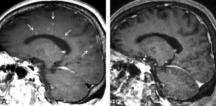
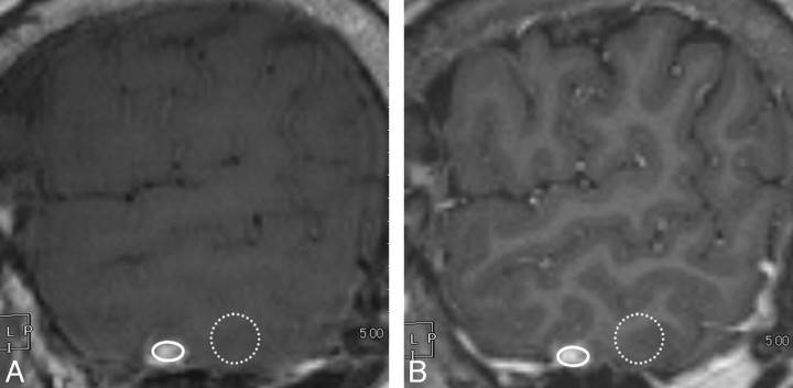
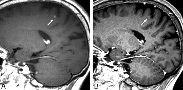
Background and purpose: Early accurate diagnosis of brain metastases is crucial for a patient's prognosis. This study aimed to compare the conspicuity and detectability of small brain metastases between contrast-enhanced 3D fast spin-echo (sampling perfection with application-optimized contrasts by using different flip angle evolutions [SPACE]) and 3D gradient-echo (GE) T1-weighted (magnetization-prepared rapid acquisition of GE [MPRAGE]) images at 3T.
Materials and methods: Sixty-nine consecutive patients with suspected brain metastases were evaluated prospectively by using SPACE and MPRAGE on a 3T MR imaging system. After careful evaluation by 2 experienced neuroradiologists, 92 lesions from 16 patients were selected as brain metastases. We compared the shorter diameter, contrast rate (CR), and contrast-to-noise ratio (CNR) of each lesion. Diagnostic ability was compared by using receiver operating characteristic (ROC) analysis. Ten radiologists (5 neuroradiologists and 5 residents) participated in the reading.
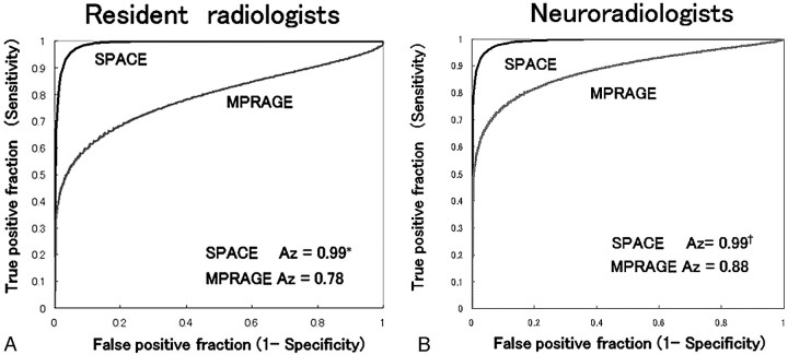
Results: The mean diameter was significantly larger by using SPACE than MPRAGE (mean, 4.5 +/- 3.7 versus 4.3 +/- 3.7 mm, P = .0014). The CR and CNR of SPACE (mean, 57.3 +/- 47.4%, 3.0 +/- 1.9, respectively) were significantly higher than those of MPRAGE (mean, 37.9 +/- 41.2%, 2.6 +/- 2.2; P < .0001, P = .04). The mean area under the ROC curve was significantly larger with SPACE than with MPRAGE (neuroradiologists, 0.99 versus 0.88, P = .013; residents, 0.99 versus 0.78, P = .0001).
Conclusions: Lesion detectability was significantly higher on SPACE than on MPRAGE, irrespective of the experience of the reader in neuroradiology. SPACE should be a promising diagnostic technique for assessing brain metastases.
Figures
References
-
- Sills AK. Current treatment approaches to surgery for brain metastases. Neurosurgery 2005. :57 (5 suppl):S24–32, discussion S1–4 - PubMed
-
- Chang EL, Hassenbusch SJ 3rd, Shiu AS, et al. The role of tumor size in the radiosurgical management of patients with ambiguous brain metastases. Neurosurgery 2003;53:272–80 - PubMed
-
- Wenz F, Hess T, Knopp MV, et al. 3D MPRAGE evaluation of lesions in the posterior cranial fossa. Magn Reson Imaging 1994;12:553–58 - PubMed
-
- Runge VM, Patel MC, Baumann SS, et al. T1-weighted imaging of the brain at 3 Tesla using a 2-dimensional spoiled gradient echo technique. Invest Radiol 2006;41:68–75 - PubMed
Publication types
MeSH terms
Substances
LinkOut - more resources
Full Text Sources
Medical