

Accelerated recovery from acute hypoxia in obese mice is due to obesity-associated up-regulation of interleukin-1 receptor antagonist
- PMID: 19213834
- PMCID: PMC2689805
- DOI: 10.1210/en.2008-1622
Accelerated recovery from acute hypoxia in obese mice is due to obesity-associated up-regulation of interleukin-1 receptor antagonist
Abstract
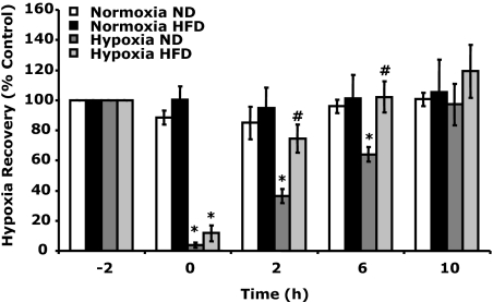
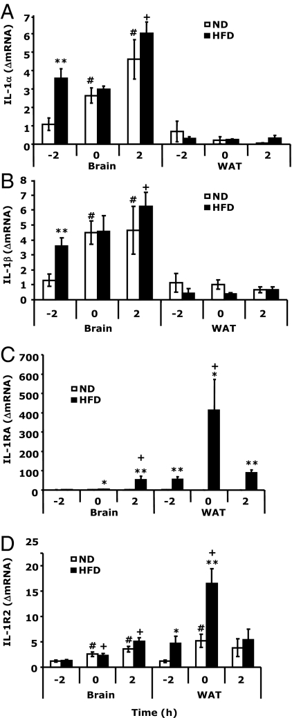
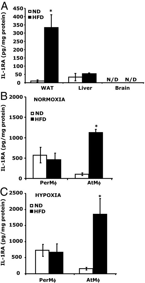
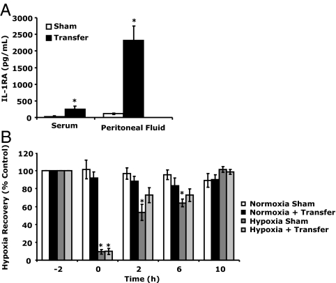
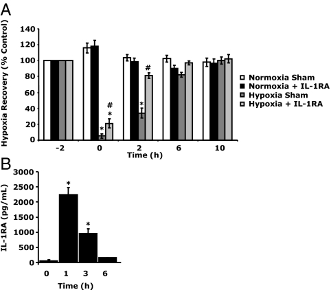
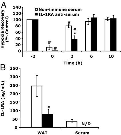
The proinflammatory consequences of obesity are thought to be due, in part, to macrophage infiltration into adipose tissue. There are, however, potential antiinflammatory consequences of obesity that include obesity-associated up-regulation of IL-1 receptor antagonist (IL-1RA). Here we show that obesity-associated up-regulation of IL-1RA speeds recovery from hypoxia. We found that high-fat diet-fed (HFD) mice recovered from acute hypoxia 5 times faster than normal-diet-fed (ND) mice. HFD mice had a 10-fold increase in serum IL-1RA when compared with ND mice. White adipose tissue (WAT) was a significant source of IL-RA, generating 330 +/- 77 pg/mg protein in HFD mice as compared with 15 +/- 5 pg/mg protein in ND mice. Peritoneal macrophages isolated from HFD mice showed little difference in IL-1RA production when compared with ND mice, but WAT macrophages from HFD mice generated 11-fold more IL-1RA than those from ND mice. When ND mice were given an ip transfer of the stromal vascular fraction portion of WAT from HFD mice, serum IL-1RA increased 836% and recovery from acute hypoxia was faster than in mice that did not receive a stromal vascular fraction transfer. To determine whether IL-1RA was important to this accelerated recovery, ND mice were administered exogenous IL-1RA prior to hypoxia, and their recovery matched that of HFD mice. Inversely, when IL-1RA was immunoabsorbed in HFD mice with IL-1RA antiserum, recovery from acute hypoxia was attenuated. Taken together these data demonstrate that HFD-induced obesity speeds recovery from hypoxia due to obesity-associated up-regulation of IL-1RA.
Figures
Similar articles
-
Interleukin-1 receptor antagonist is upregulated during diet-induced obesity and regulates insulin sensitivity in rodents.Diabetologia. 2006 Feb;49(2):387-93. doi: 10.1007/s00125-005-0046-x. Epub 2005 Dec 30. Diabetologia. 2006. PMID: 16385385
-
Behavioral recovery from acute hypoxia is reliant on leptin.Brain Behav Immun. 2009 Feb;23(2):169-75. doi: 10.1016/j.bbi.2008.09.011. Epub 2008 Sep 27. Brain Behav Immun. 2009. PMID: 18854211 Free PMC article.
-
Adipose tissue is a major source of interleukin-1 receptor antagonist: upregulation in obesity and inflammation.Diabetes. 2003 May;52(5):1104-10. doi: 10.2337/diabetes.52.5.1104. Diabetes. 2003. PMID: 12716739
-
Exercise training inhibits inflammation in adipose tissue via both suppression of macrophage infiltration and acceleration of phenotypic switching from M1 to M2 macrophages in high-fat-diet-induced obese mice.Exerc Immunol Rev. 2010;16:105-18. Exerc Immunol Rev. 2010. PMID: 20839495
-
Dynamic, M2-like remodeling phenotypes of CD11c+ adipose tissue macrophages during high-fat diet--induced obesity in mice.Diabetes. 2010 May;59(5):1171-81. doi: 10.2337/db09-1402. Epub 2010 Feb 25. Diabetes. 2010. PMID: 20185806 Free PMC article.
Cited by
-
Sickness behavior induced by endotoxin can be mitigated by the dietary soluble fiber, pectin, through up-regulation of IL-4 and Th2 polarization.Brain Behav Immun. 2010 May;24(4):631-40. doi: 10.1016/j.bbi.2010.01.015. Epub 2010 Feb 6. Brain Behav Immun. 2010. PMID: 20138982 Free PMC article.
-
The biobehavioral and neuroimmune impact of low-dose ionizing radiation.Brain Behav Immun. 2012 Feb;26(2):218-27. doi: 10.1016/j.bbi.2011.09.006. Epub 2011 Sep 21. Brain Behav Immun. 2012. PMID: 21958477 Free PMC article.
-
Fasting induces an anti-inflammatory effect on the neuroimmune system which a high-fat diet prevents.Obesity (Silver Spring). 2011 Aug;19(8):1586-94. doi: 10.1038/oby.2011.73. Epub 2011 Apr 28. Obesity (Silver Spring). 2011. PMID: 21527899 Free PMC article.
-
Fasting Induces IL-1 Resistance and Free-Fatty Acid-Mediated Up-Regulation of IL-1R2 and IL-1RA.Front Immunol. 2014 Jul 9;5:315. doi: 10.3389/fimmu.2014.00315. eCollection 2014. Front Immunol. 2014. PMID: 25071776 Free PMC article.
-
Individually ventilated cages cause chronic low-grade hypoxia impacting mice hematologically and behaviorally.Brain Behav Immun. 2012 Aug;26(6):951-8. doi: 10.1016/j.bbi.2012.04.008. Epub 2012 May 3. Brain Behav Immun. 2012. PMID: 22561683 Free PMC article.
References
-
- Lago F, Dieguez C, Gómez-Reino J, Gualillo O 2007 Adipokines as emerging mediators of immune response and inflammation. Nat Clin Pract Rheumatol 3:716–724 - PubMed
-
- Kershaw EE, Flier JS 2004 Adipose tissue as an endocrine organ. J Clin Endocrinol Metab 89:2548–2556 - PubMed
-
- Hauner H 2005 Secretory factors from human adipose tissue and their functional role. Proc Nutr Soc 64:163–169 - PubMed
-
- Fain JN, Madan AK, Hiler ML, Cheema P, Bahouth SW 2004 Comparison of the release of adipokines by adipose tissue, adipose tissue matrix, and adipocytes from visceral and subcutaneous abdominal adipose tissues of obese humans. Endocrinology 145:2273–2282 - PubMed
-
- Meliga E, Strem BM, Duckers HJ, Serruys PW 2007 Adipose-derived cells. Cell Transplant 16:963–970 - PubMed
Publication types
MeSH terms
Substances
Grants and funding
LinkOut - more resources
Full Text Sources
Medical
Research Materials
