. 2009 Feb;5(2):e1000293.
doi: 10.1371/journal.ppat.1000293. Epub 2009 Feb 13.

Detailed analysis of sequence changes occurring during vlsE antigenic variation in the mouse model of Borrelia burgdorferi infection

Affiliations

Detailed analysis of sequence changes occurring during vlsE antigenic variation in the mouse model of Borrelia burgdorferi infection

Loïc Coutte et al. PLoS Pathog. 2009 Feb.

Abstract

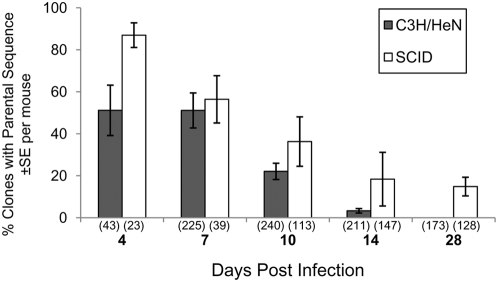
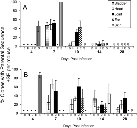
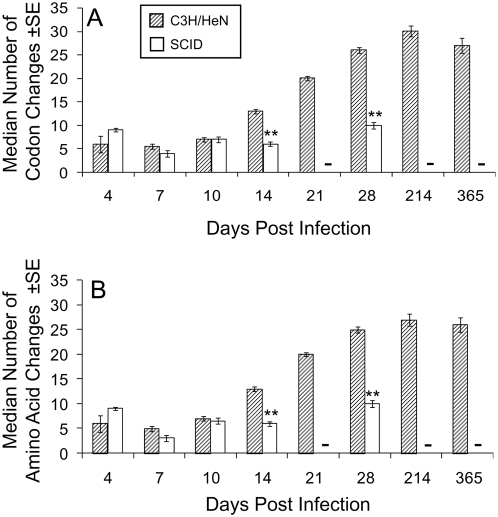
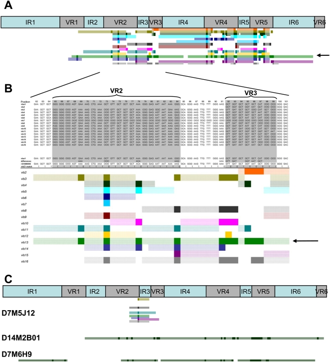
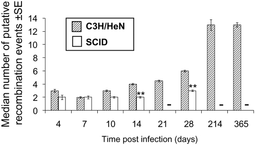
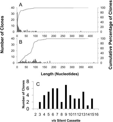
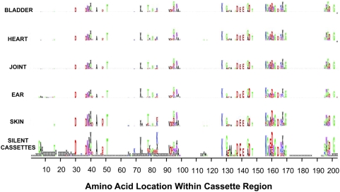
Lyme disease Borrelia can infect humans and animals for months to years, despite the presence of an active host immune response. The vls antigenic variation system, which expresses the surface-exposed lipoprotein VlsE, plays a major role in B. burgdorferi immune evasion. Gene conversion between vls silent cassettes and the vlsE expression site occurs at high frequency during mammalian infection, resulting in sequence variation in the VlsE product. In this study, we examined vlsE sequence variation in B. burgdorferi B31 during mouse infection by analyzing 1,399 clones isolated from bladder, heart, joint, ear, and skin tissues of mice infected for 4 to 365 days. The median number of codon changes increased progressively in C3H/HeN mice from 4 to 28 days post infection, and no clones retained the parental vlsE sequence at 28 days. In contrast, the decrease in the number of clones with the parental vlsE sequence and the increase in the number of sequence changes occurred more gradually in severe combined immunodeficiency (SCID) mice. Clones containing a stop codon were isolated, indicating that continuous expression of full-length VlsE is not required for survival in vivo; also, these clones continued to undergo vlsE recombination. Analysis of clones with apparent single recombination events indicated that recombinations into vlsE are nonselective with regard to the silent cassette utilized, as well as the length and location of the recombination event. Sequence changes as small as one base pair were common. Fifteen percent of recovered vlsE variants contained "template-independent" sequence changes, which clustered in the variable regions of vlsE. We hypothesize that the increased frequency and complexity of vlsE sequence changes observed in clones recovered from immunocompetent mice (as compared with SCID mice) is due to rapid clearance of relatively invariant clones by variable region-specific anti-VlsE antibody responses.


Conflict of interest statement

The authors have declared that no competing interests exist.

Figures

Figure 1
Figure 1. B. burgdorferi clones having the parental vlsE sequence are cleared more rapidly during infection of immunocompetent C3H/HeN mice than in immunodeficient SCID mice.
The numbers in parentheses represent the total number of clones at each time point.
Figure 2
Figure 2. Relative persistence of clones retaining the parental vlsE sequence at different tissue sites during the course of infection of C3H/HeN and SCID mice.
Panel (A) represents data obtained from C3H/HeN mice, whereas Panel (B) contains data from SCID mice. Results for each time point are presented in the order shown (bladder, heart, joint, ear, and skin). - No data available (no positive culture obtained, or culture not done). 0 No parental sequences identified for that tissue and timepoint.
Figure 3
Figure 3. Median number of vlsE codon changes and predicted amino acid changes in variants during the time course of infection of C3H/HeN or SCID mice.
Clones with the parental vlsE sequence were excluded from this analysis. (A) vlsE codon changes. (B) Predicted amino acid changes. ** indicates a significant difference (P<0.01) between organisms from C3H/HeN and SCID mice at the time points indicated, as calculated by unpaired Student's t test. - No data available (culture not done).
Figure 4
Figure 4. Schematic representation of vlsE recombination patterns.
(A) Upper panel represents the locations of the 6 invariable regions (IR) and the 6 variable regions (VR) within the vlsE cassette region. Lower panel illustrates the pattern of recombination of clone D10M8H8 showing sequence changes between VR1 to VR5. (B) Magnified region of D10M8H8 recombination pattern. The top portion of the diagram shows the alignment between the parental vlsE sequence (vlsE1), the vls silent cassettes vls2 through vls16, and the vlsE variant. The line “differences” highlights the difference between the vlsE and the variant sequences. In the lower panel, each colored line schematically represents regions of the 15 silent cassette sequences which could be involved in the variant sequence changes. Each dark colored block represents a region of sequence change within the variant sequence that is present in the selected silent cassette sequence. The light colored regions in each line represent segments adjacent to sequence changes that are identical in vlsE1, the variant and the selected silent cassette sequence (i.e. the maximal possible recombination region). From this analysis, two likely recombination events using silent cassette vls13 (green) as template were identified: from VR1 to VR3 (arrow, panel B) and from VR4 to VR5 (see panel A). (C) Recombination patterns obtained for variant sequences, exemplifying the following patterns: a single codon change involving any one of several possible silent cassettes (D7M5J12), a long recombination event with silent cassette 8 spanning VR2 through VR6 (D14M2B01), and 4 intermittent recombination events involving silent cassette 13 (D7M6H9).
Figure 5
Figure 5. Progressive recombination in vlsE variant clones.
(A) Schematic representation of possible recombination events for three clones derived from a single day 10 bladder culture. The shaded gray boxes indicate the variable regions (VR). Colored bars represent the maximum possible length of DNA involved in a putative recombination event for each vls silent cassette. Dark colored blocks within the colored bars represent observed sequence changes between vlsE and the variant sequence that are present in the selected silent cassette. (B) Postulated sequence of recombination events leading to the three clones.
Figure 6
Figure 6. Median number of putative recombination events in variant B. burgdorferi clones during the time course of infection.
Hatched and empty bars represent the populations of bacteria recovered from C3H/HeN or SCID mice, respectively. ** indicates a significant difference (P<0.01) between the results obtained for C3H/HeN and SCID mice for that time point, as calculated by unpaired Student's t test.
Figure 7
Figure 7. Lengths of minimum and maximum predicted recombination events in 126 clones identified as having a single, well-defined recombination event.
Histograms of the deduced (A) minimum and (B) maximum lengths of recombination are depicted as bars; the cumulative percentage of clones having predicted minimal or maximal recombination lengths≤the length shown are represented as lines. Panel C represents the putative silent cassette usage in clones with a single recombination event in which the silent cassette source could be determined unambiguously.
Figure 8
Figure 8. The locations and amino acid utilization of deduced VlsE amino acid changes paralleled the changes predicted by the sequence differences between vlsE1 and the silent cassettes.
The distribution of amino acid changes found in the variant sequences from different infected tissues were depicted as sequence logo patterns using the program WebLogo . The height of the letter is proportional to the frequency of changes. The letter “x” indicates a 3-nt indel, and the asterisk a stop codon. The panels for bladder, heart, joint, ear, and skin represent the observed changes in the variant sequences recovered from the respective tissue at all time points during B. burgdorferi infection of C3H/HeN or SCID mice. The silent cassette panel represents the relative probability of a given amino acid change at each position of vlsE1, based on the amino acids encoded by the silent cassette sequences at each position in the vlsE1/silent cassette alignment.

References

    1. Steere AC, Coburn J, Glickstein L. The emergence of Lyme disease. J Clin Invest. 2004;113:1093–1101. - PMC - PubMed
    1. Craft JE, Grodzicki RL, Shrestha M, Fischer DK, Garcia-Blanco M, et al. The antibody response in Lyme disease. Yale J Biol Med. 1984;57:561–565. - PMC - PubMed
    1. Craft JE, Grodzicki RL, Steere AC. Antibody response in Lyme disease: evaluation of diagnostic tests. J Infect Dis. 1984;149:789–795. - PubMed
    1. Dressler F, Whalen JA, Reinhardt BN, Steere AC. Western blotting in the serodiagnosis of Lyme disease. J Infect Dis. 1993;167:392–400. - PubMed
    1. Feder HM, Jr, Gerber MA, Luger SW, Ryan RW. Persistence of serum antibodies to Borrelia burgdorferi in patients treated for Lyme disease. Clin Infect Dis. 1992;15:788–793. - PubMed

Publication types

MeSH terms