. 2009 Feb;5(2):e1000382.
doi: 10.1371/journal.pgen.1000382. Epub 2009 Feb 13.

Disease-associated mutant ubiquitin causes proteasomal impairment and enhances the toxicity of protein aggregates

Affiliations

Disease-associated mutant ubiquitin causes proteasomal impairment and enhances the toxicity of protein aggregates

Elizabeth M H Tank et al. PLoS Genet. 2009 Feb.

Abstract

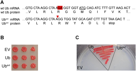
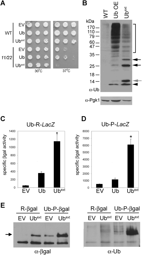
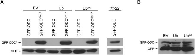
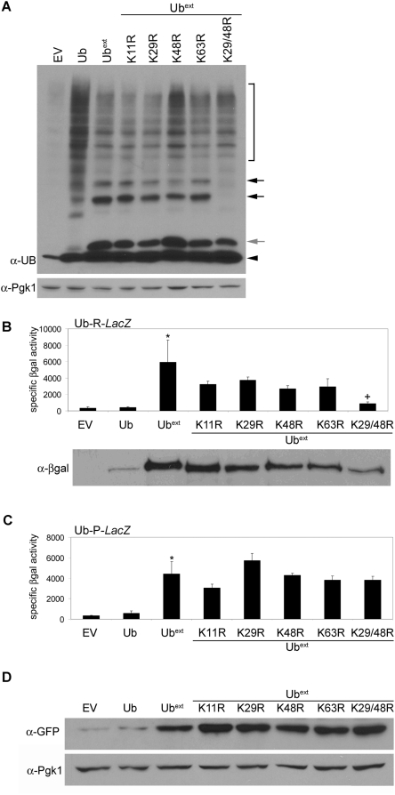
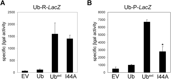
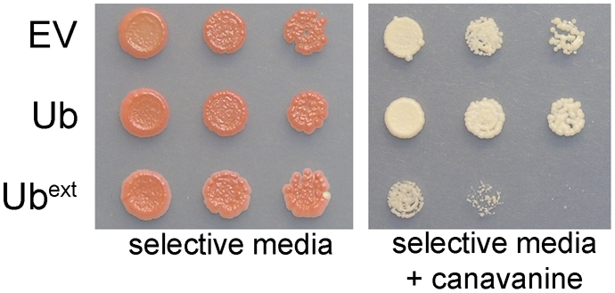
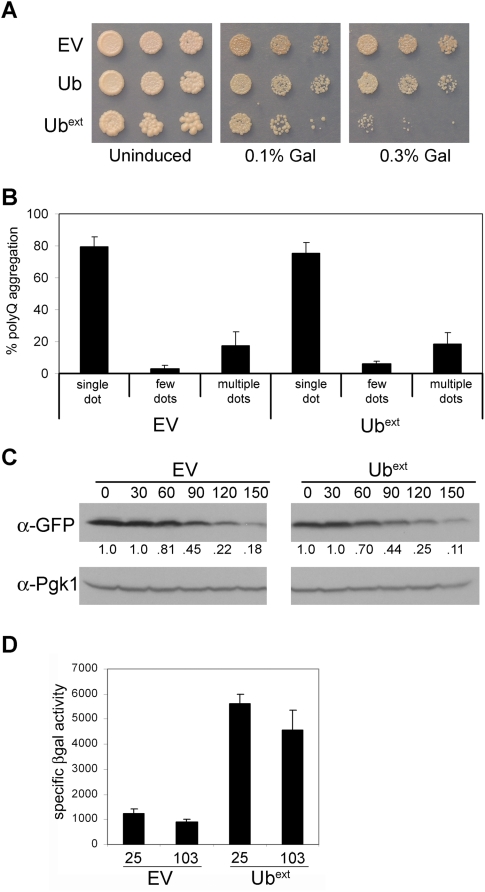
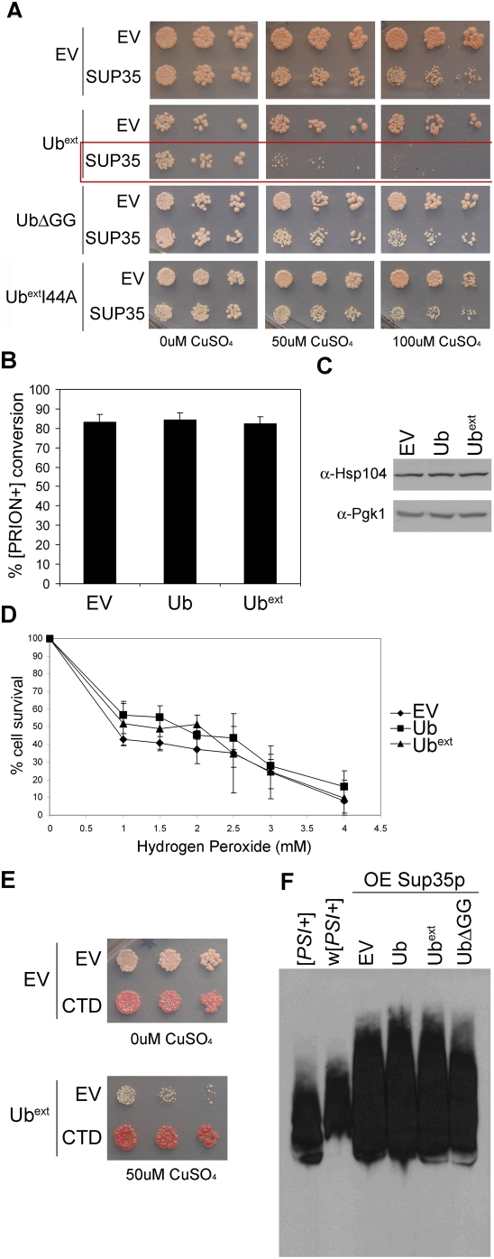
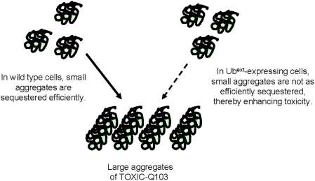
Protein homeostasis is critical for cellular survival and its dysregulation has been implicated in Alzheimer's disease (AD) and other neurodegenerative disorders. Despite the growing appreciation of the pathogenic mechanisms involved in familial forms of AD, much less is known about the sporadic cases. Aggregates found in both familial and sporadic AD often include proteins other than those typically associated with the disease. One such protein is a mutant form of ubiquitin, UBB+1, a frameshift product generated by molecular misreading of a wild-type ubiquitin gene. UBB+1 has been associated with multiple disorders. UBB+1 cannot function as a ubiquitin molecule, and it is itself a substrate for degradation by the ubiquitin/proteasome system (UPS). Accumulation of UBB+1 impairs the proteasome system and enhances toxic protein aggregation, ultimately resulting in cell death. Here, we describe a novel model system to investigate how UBB+1 impairs UPS function and whether it plays a causal role in protein aggregation. We expressed a protein analogous to UBB+1 in yeast (Ub(ext)) and demonstrated that it caused UPS impairment. Blocking ubiquitination of Ub(ext) or weakening its interactions with other ubiquitin-processing proteins reduced the UPS impairment. Expression of Ub(ext) altered the conjugation of wild-type ubiquitin to a UPS substrate. The expression of Ub(ext) markedly enhanced cellular susceptibility to toxic protein aggregates but, surprisingly, did not induce or alter nontoxic protein aggregates in yeast. Taken together, these results suggest that Ub(ext) interacts with more than one protein to elicit impairment of the UPS and affect protein aggregate toxicity. Furthermore, we suggest a model whereby chronic UPS impairment could inflict deleterious consequences on proper protein aggregate sequestration.


Conflict of interest statement

The authors have declared that no competing interests exist.

Figures

Figure 1
Figure 1. Ubext does not function as ubiquitin.
(A) A schematic diagram depicting the wild type and mutant ubiquitin (Ubext) mRNA and protein sequences beginning at nucleotide 207 of UBI4. The underlined ATG denotes the beginning of the next ubiquitin open reading frame in the tandem array. The red triangle signifies the site of the dinucleotide GA deletion. (B) Ubext expression does not affect logarithmically growing yeast. Serial dilutions of wild type yeast ectopically expressing Ubext, excess wild type ubiquitin (Ub), or an empty vector (EV) control were spotted onto selective medium. (C) Ubext does not behave as wild type ubiquitin and cannot compensate for the loss of UBI4. The Δubi4 strain was transformed with EV, Ub, and Ubext. Transformants were plated onto selective medium following growth into stationary phase.
Figure 2
Figure 2. Expression of Ubext causes proteasomal impairment.
(A) Ubext displays synthetic lethality with proteasome mutants. Wild type (WT) and temperature-sensitive proteasome mutant cells, pre1-1 pre2-2 (11/22), were transformed with plasmids containing empty vector (EV), ubiquitin (Ub) and Ubext. Serial dilutions of cells were spotted onto selective medium and grown at 30°C and 37°C. (B) Cells expressing Ubext show a distinct pattern of ubiquitin conjugation. Protein lysate from wild type yeast cells containing an empty vector (WT), extra ubiquitin (Ub OE), or Ubext were analyzed by SDS-PAGE and western blot using an anti-ubiquitin antibody. Ubext causes an increase in ubiquitin-conjugated proteins (bracket) as compared to WT. The black arrowhead indicates ubiquitin monomer. The grey arrow points to Ubext. Black arrows represent conjugated Ubext. Pgk1p expression was probed to assess protein loading on the membrane. (C) Ubext-expressing cells impair the degradation of the N-end rule substrate R-β-galactosidase (βgal). Cells containing EV, Ub, or Ubext were transformed with pGal-Ub-R-LacZ. The stability of R-βgal was measured by specific activity (luminescence units/µg protein). The asterisk (*) indicates statistical significance between wild type Ub and Ubext (p = 0.0013). (D) Ubext-expression prevents the efficient proteasomal degradation of a ubiquitin fusion degradation substrate. The stability of Ub-P-LacZ was evaluated as in B. The asterisk (*) indicates statistical significance between wild type Ub and Ubext (p = 0.0005). (E) Ubiquitinated reporter substrates are present in Ubext-expressing cells. Wild type cells containing the Ub-X-LacZ reporter constructs and expressing Ubext or the control (EV) were analyzed for ubiquitinated βgal protein. βgal protein was immunoprecipitated with an anti-βgal antibody (left) and the bound fractions were blotted with an anti-ubiquitin antibody (right). The arrow indicates full length βgal protein.
Figure 3
Figure 3. Ubext-expressing cells can degrade a ubiquitin-independent substrate.
(A) Expression of Ubext does not impair the degradation of GFP-ODC. Cells containing a stable GFP construct were transformed with empty vector (EV), Ub, or Ubext. These cells were then transformed with plasmids expressing either GFP-ODC or GFP-ODCC441A. A proteasome mutant strain (pre1-1 pre2-2, abbreviated 11/22) was transformed with both the GFP and GFP-ODC constructs and as expected, both were stable. Protein lysates were separated by SDS-PAGE and analyzed by western blot using an anti-GFP antibody. GFP-ODC* denotes either GFP-ODC or GFP-ODCC441A. (B) Ubext-expressing cells accumulate approximately an equal amount GFP-ODC in comparison to controls. Protein lysates from cells containing EV, Ub or Ubext co-expressing GFP-ODC were analyzed by SDS-PAGE and western blot with an anti-GFP antibody and visualized after prolonged exposure (1 hour).
Figure 4
Figure 4. Ubiquitin conjugation of Ubext is required for stabilization of N-end rule but not UFD substrates.
(A) Lysines 29 and 48 are required for ubiquitin conjugation to Ubext. Protein lysate from cells containing empty vector (EV), Ub, Ubext, or UbextKxR were analyzed by SDS-PAGE and western blot using an anti-ubiquitin antibody. The black arrowhead indicates mono-ubiquitin. The grey arrow points to Ubext. Black arrows represent conjugated Ubext. Pgk1p was probed to assess protein loading (lower). (B) The conjugation of Ubext is necessary for the impaired degradation of the N-end rule substrate R-βgal. Cells containing EV, Ub, Ubext, or UbextKxR mutants were transformed with pGalUb-R-LacZ and analyzed by βgal activity assay. The asterisk (*) denotes statistical significance between Ubext and EV (p = 0.0239). The cross (+) indicates statistical significance between Ubext and UbextK29/48R (p = 0.032). There is no statistically significant difference between Ubext and UbextK11R (p = 0.1602). Lower: Corresponding βgal protein levels from the lysates used in the βgal activity assay were detected by SDS-PAGE and western blot using an anti-βgal antibody. (C) Ubiquitin conjugation of Ubext is not necessary for the impaired degradation of the UFD substrate Ub-P-βgal. Cells containing EV, Ub, Ubext, or UbextKxR mutants were transformed with pGalUb-P-LacZ and analyzed by βgal activity assay. The asterisk (*) denotes statistical significance between Ubext and EV (p = 0.0055). There is no statistically significant difference between Ubext and UbextK48/29R (p = 0.4558). (D) Ubext-ubiquitin conjugation is not necessary to impair the degradation of a second UFD substrate, UbG76V-GFP. Cells containing EV, Ub, Ubext, or UbextKxR mutants were transformed with UbG76V-GFP and analyzed by SDS-PAGE and western blot using an anti-GFP antibody. The blot was reprobed for Pgk1p as a loading control.
Figure 5
Figure 5. Mutation of the Ubext hydrophobic patch (I44A) moderately affects proteasomal impairment.
(A) UbextI44A still inhibits N-end rule substrate degradation. Cells containing pGal-Ub-R-LacZ were transformed with empty vector (EV), Ub, Ubext or UbextI44A (I44A) and the stability of R-βgal was measured by βgal activity assay. (B) UbextI44A moderately enhances the degradation of a UFD substrate. Cells containing pGal-Ub-P-LacZ were transformed with EV, Ub, Ubext or UbextI44A (I44A) and the stability of Ub-P-βgal was measured by βgal activity assay. The asterisk (*) indicates statistical significance between Ubext and UbextI44A (p = 0.0007).
Figure 6
Figure 6. Ubext expression increases cellular sensitivity to misfolded proteins.
Ubext-expressing cells cannot tolerate excess misfolded proteins generated by the incorporation of canavanine. Serial dilutions of cells containing EV, Ub, or Ubext were spotted onto selective medium and selective medium containing 400 µM canavanine.
Figure 7
Figure 7. Ubext expression does enhance the toxicity of polyglutamine expanded protein but does not affect protein aggregation.
(A) Cells containing empty vector (EV), Ub, or Ubext were transformed with a galactose-inducible TOXIC-Q103 construct, which induces cell death in the presence of galactose. Serial dilutions of transformants were spotted onto selective medium (uninduced) and selective media containing either 0.1% or 0.3% galactose. (B) Pre-existing non-toxic HttQ103-GFP aggregates were not altered in the presence of Ubext. Cells transformed with non-toxic HttQ103-GFP and EV or Ubext were analyzed by fluorescence microscopy. The abundance and pattern of aggregates (dots) was evaluated as described in Materials and Methods using three independent cultures for each sample. Data are expressed as a percentage of the total cells containing aggregates. (C) TOXIC-Q103 protein is not stabilized in the presence of Ubext. Cells expressing TOXIC-Q103 in the absence (EV) or presence of Ubext were treated with cycloheximide and harvested at the indicated times post translational shut off (in minutes). Cell lysates were analyzed by western blot for the expression of TOXIC-Q103, (which is CFP tagged) using an anti-GFP antibody. Relative protein abundance was quantified as a ratio of the total (below). The membrane was reprobed with an anti-Pgk1 antibody to show protein loading. (D) TOXIC-Q103 protein aggregates do not cause proteasomal impairment. pGal-Ub-P-LacZ containing cells with and without Ubext were transformed with galactose-inducible Q25 or TOXIC-Q103 constructs. The transformants were grown in selective medium containing galactose for 24 hours and the stability of the Ub-P-βgal substrate was measured by βgal activity assay.
Figure 8
Figure 8. Expression of Ubext enhances the susceptibility of cells to toxic Sup35p aggregates but does not affect Sup35p aggregation.
(A) [PSI+] cells expressing Ubext show reduced cell viability with lower induction of Sup35p. [PSI+] cells containing empty vector (EV), Ubext, UbΔGG, or UbextI44A were transformed with a copper-inducible SUP35 or EV and analyzed for growth by spotting serial dilutions onto selective media containing 0, 50, or 100 µM CuSO4. At 300 µM CuSO4, [PSI+] cells over expressing Sup35p alone are not viable (not shown). (B) Prion conversion or induction was not enhanced in cells expressing Ubext. [psi−] cells expressing pSup35 or the control (EV) were transformed with empty vector (EV), Ub or Ubext and were analyzed for [PSI+] prion formation by monitoring colony color (the appearance of pink colonies). The graph represents the average of three independent cultures in which approximately 2,000 colonies per culture were evaluated for conversion. (C) Hsp104 protein levels are not enhanced in Ubext-expressing cells. Protein lysate from cells containing EV, Ub, or Ubext were subject to SDS-PAGE and western blot using an anti-Hsp104 antibody. Pgk1p expression was analyzed as a loading control. (D) The expression of Ubext did not alter cell survival in the presence of oxidative stress. Cells containing EV, Ub, or Ubext were treated with increasing concentrations of hydrogen peroxide (H2O2) and the number of viable cells was graphed as a percentage of the untreated. (E) The C-terminal domain of Sup35p (CTD) rescued the enhance susceptibility caused by Ubext in [PSI+] cells over expressing Sup35p. Upper: [PSI+]-mediated nonsense suppression is alleviated by expression of the CTD. [PSI+] cells containing EV show more nonsense suppression (the colony color is light pink). However, [PSI+] cells expressing the CTD display efficient translation termination and the colonies are red. Lower: [PSI+] cells expressing Ubext in addition to excess Sup35p (induced by 50 µM copper) are rescued from death by the expression of the CTD. (F) Sup35 protein aggregates were not altered by the presence of Ubext. Sup35p aggregates in strong [PSI+] ([PSI+]) and a weak strain variant of [PSI+] (w[PSI+]) were analyzed by SDD-AGE. The difference in Sup35p aggregate size of these prion strain variants can be appreciated by this method (compare [PSI+] to w[PSI+]). Sup35p aggregates from cells expressing excess Sup35p (OE Sup35p) and expressing Ub, Ubext, UbΔGG or containing an EV control were analyzed by SDD-AGE and western blot with an anti-Sup35 antibody.
Figure 9
Figure 9. Model for Ubext affects on toxic protein aggregates.
We propose that enhanced protein aggregate toxicity in Ubext-expressing cells is due to the inability of misfolded amyloidogenic proteins to be properly sequestered. The small soluble oligomers are more toxic than the large insoluble protein aggregates. UPS impairment caused by the expression of Ubext may hinder the rapid sequestration or retention of toxic oligomers into large protein aggregates.
Figure 10
Figure 10. Protein aggregate toxicity is enhanced by perturbations of the UPS and protein aggregate solubility is enhanced by Ubext.
(A) Limiting ubiquitination also decreases cellular survival in the presence of TOXIC-Q103. Cellular viability of TOXIC-Q103 (103) or the Q25 control expressed in ts uba1-204 cells was measured at the permissive temperature (30°C) and a restrictive temperature (32°C). The graph represents the percentage of viable cells from the inducing plates compared to cells grown on non-inducing medium. The asterisk (*) indicates statistical significance between 25 and 103 at 30°C (p = 0.0007), the cross (+) indicates statistical significance between 25 and 103 at 32°C (p = 0.0003), and the double asterisk (**) indicates statistical significance between 103 at 30°C and 32°C (p = 0.0068). (B) Increasing misfolded proteins enhanced toxicity in the presence of TOXIC-Q103. Cell expressing TOXIC-PQ103 (103) and the Q25 control (25) were spotted onto inducing medium and inducing medium containing 200 µM canavanine (Can). (C) The cellular susceptibility of over expressed Sup35p in [PSI+] cells in the presence of canavanine is not as detrimental as the co-expression of Ubext. Cells expressing excess Sup35p (induced with 200 µM CuSO4) were spotted onto plates containing 400 µM canavanine (Can). Sup35 over expressing cells are slightly less viable in the presence of 400 µM canavanine. All cells died at higher concentrations of CuSO4 and canavanine. (D) Cells expressing Ubext contain more soluble TOXIC-Q103 protein. Cells expressing TOXIC-Q103 in the presence of Ubext or absence (EV) were lysed and the soluble protein was analyzed by western blot after high speed ultracentrifugation. Densitometry was performed to determine the amount of soluble TOXIC-Q103 protein normalized to the total protein for each sample and graphed in relative arbitrary units. Three independent cultures for each sample were analyzed. The asterisk (*) denotes statistical significance (p = 0.0052).
Figure 11
Figure 11. Ubext alters the ubiquitination pattern of a UPS substrate.
(A) R-βgal ubiquitination pattern is not altered in cells expressing Ubext. pGalUb-R-LacZ was transformed into proteasome mutant cells (pre1-1 pre2-2) expressing Ubext or EV and R-βgal was analyzed by immunoprecipitation (IP). Membranes were probed with anti-βgal and anti-ubiquitin antibodies. Arrow indicates full length βgal protein. (B) Ub-P-βgal ubiquitination is affected in cells expressing Ubext. Ub-P-βgal IPs were performed as in A. A subtle but reproducible difference in ubiquitination pattern was observed. Three independent IPs are shown. Arrowheads highlight distinct bands present in the EV lanes that are absent in Ubext lanes.

References

    1. Goedert M, Spillantini MG. A century of Alzheimer's disease. Science. 2006;314:777–781. - PubMed
    1. Jentsch S. The ubiquitin-conjugation system. Annu Rev Genet. 1992;26:179–207. - PubMed
    1. Johnson ES, Bartel B, Seufert W, Varshavsky A. Ubiquitin as a degradation signal. Embo J. 1992;11:497–505. - PMC - PubMed
    1. Varshavsky A. The ubiquitin system. Trends Biochem Sci. 1997;22:383–387. - PubMed
    1. Schwartz AL, Ciechanover A. The ubiquitin-proteasome pathway and pathogenesis of human diseases. Annu Rev Med. 1999;50:57–74. - PubMed

Publication types

MeSH terms