

Oxygen as modulator of trophoblast invasion
- PMID: 19215320
- PMCID: PMC2714634
- DOI: 10.1111/j.1469-7580.2008.01036.x
Oxygen as modulator of trophoblast invasion
Abstract
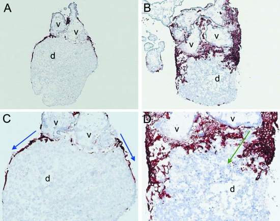
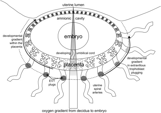
At the time of blastocyst implantation the uterine spiral arteries have already undergone morphological changes in the absence of any extravillous trophoblast invasion. Only 2 weeks after implantation, extravillous trophoblast cells develop and come into first contact with decidual tissues. Invading through the decidual interstitium, extravillous trophoblasts potentially reach and transform spiral arteries into uteroplacental arteries. Spiral arterial erosion starts at about mid-first trimester, whereas flow of maternal blood into the intervillous space is continuously established only at the beginning of the second trimester. One key regulator of the number of extravillous trophoblasts is oxygen. The steep gradient in oxygen concentration within the first trimester placenta is diminished with the onset of maternal blood flow. This gradient is used by the trophoblast to generate a large number of invasive cells to adapt the arterial vasculature in the placental bed to the growing needs of the fetus. Changes in oxygen concentrations or other factors leading to alterations in the rates of proliferation and/or apoptosis of extravillous trophoblast clearly impact on the remodelling of the vessels. The respective consequences of a failure in trophoblast invasion are growth restrictions of the baby and perhaps other pregnancy complications.
Figures
References
-
- Aplin JD. Implantation, trophoblast differentiation and haemochorial placentation: mechanistic evidence in vivo and in vitro. J Cell Sci. 1991;99:681–692. - PubMed
-
- Benirschke K, Kaufmann P, Baergen R. Pathology of the Human Placenta. 5th edn. New York: Springer; 2006.
-
- Boyd JD, Hamilton WJ. Cells in the spiral arteries of the pregnant uterus. J Anat. 1956;90:595.
-
- Boyd JD, Hamilton WJ. Development and structure of the human placenta from the end of the 3rd month of gestation. J Obstet Gynaecol Br Commonw. 1967;74:161–226. - PubMed
-
- Brettner A. Zum Verhalten der sekundaeren Wand der Uteroplacentargefaesse bei der decidualen Reaktion. Acta Anat. 1964;57:367–376. - PubMed
Publication types
MeSH terms
LinkOut - more resources
Full Text Sources
