

Mutations in STIL, encoding a pericentriolar and centrosomal protein, cause primary microcephaly
- PMID: 19215732
- PMCID: PMC2668020
- DOI: 10.1016/j.ajhg.2009.01.017
Mutations in STIL, encoding a pericentriolar and centrosomal protein, cause primary microcephaly
Abstract
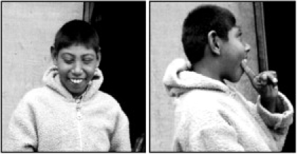
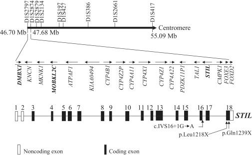
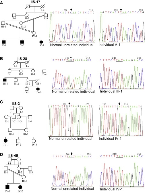
Primary microcephaly (MCPH) is an autosomal-recessive congenital disorder characterized by smaller-than-normal brain size and mental retardation. MCPH is genetically heterogeneous with six known loci: MCPH1-MCPH6. We report mapping of a novel locus, MCPH7, to chromosome 1p32.3-p33 between markers D1S2797 and D1S417, corresponding to a physical distance of 8.39 Mb. Heterogeneity analysis of 24 families previously excluded from linkage to the six known MCPH loci suggested linkage of five families (20.83%) to the MCPH7 locus. In addition, four families were excluded from linkage to the MCPH7 locus as well as all of the six previously known loci, whereas the remaining 15 families could not be conclusively excluded or included. The combined maximum two-point LOD score for the linked families was 5.96 at marker D1S386 at theta = 0.0. The combined multipoint LOD score was 6.97 between markers D1S2797 and D1S417. Previously, mutations in four genes, MCPH1, CDK5RAP2, ASPM, and CENPJ, that code for centrosomal proteins have been shown to cause this disorder. Three different homozygous mutations in STIL, which codes for a pericentriolar and centrosomal protein, were identified in patients from three of the five families linked to the MCPH7 locus; all are predicted to truncate the STIL protein. Further, another recently ascertained family was homozygous for the same mutation as one of the original families. There was no evidence for a common haplotype. These results suggest that the centrosome and its associated structures are important in the control of neurogenesis in the developing human brain.
Figures
Comment in
-
STIL on my small brain: a new gene for microcephaly.Clin Genet. 2009 Dec;76(6):501-2. doi: 10.1111/j.1399-0004.2009.01297.x. Clin Genet. 2009. PMID: 19930152 No abstract available.
References
-
- Kumar A., Blanton S.H., Babu M., Markandaya M., Girimaji S.C. Genetic analysis of primary microcephaly in Indian families: Novel ASPM mutations. Clin. Genet. 2004;66:341–348. - PubMed
-
- Bond J., Roberts E., Mochida G.H., Hampshire D.J., Scott S., Askham J.M., Springell K., Mahadevan M., Crow Y.J., Markham A.F. ASPM is a major determinant of cerebral cortical size. Nat. Genet. 2002;32:316–320. - PubMed
-
- Cowie V. The genetics and sub-classification of microcephaly. J. Ment. Defic. Res. 1960;4:42–47. - PubMed
Publication types
MeSH terms
Substances
LinkOut - more resources
Full Text Sources
Other Literature Sources
Molecular Biology Databases
Miscellaneous
