

Variability of target and normal structure delineation for breast cancer radiotherapy: an RTOG Multi-Institutional and Multiobserver Study
- PMID: 19215827
- PMCID: PMC2911777
- DOI: 10.1016/j.ijrobp.2008.10.034
Variability of target and normal structure delineation for breast cancer radiotherapy: an RTOG Multi-Institutional and Multiobserver Study
Abstract
Purpose: To quantify the multi-institutional and multiobserver variability of target and organ-at-risk (OAR) delineation for breast-cancer radiotherapy (RT) and its dosimetric impact as the first step of a Radiation Therapy Oncology Group effort to establish a breast cancer atlas.
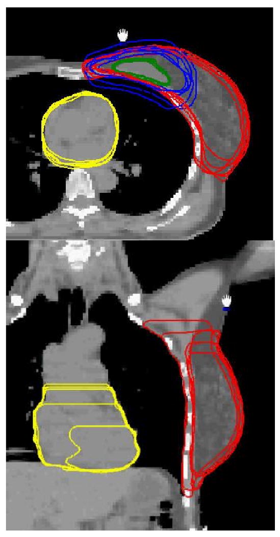
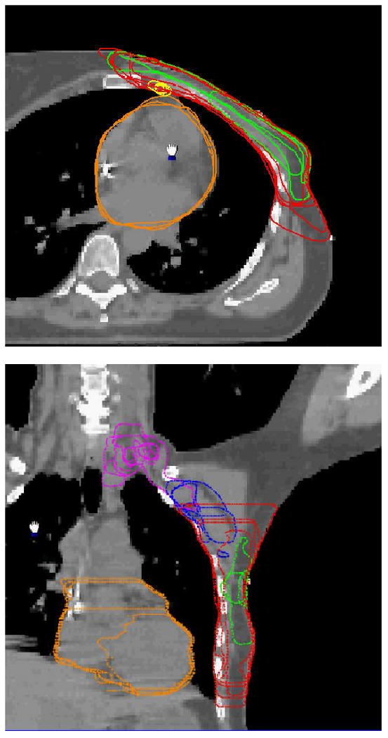
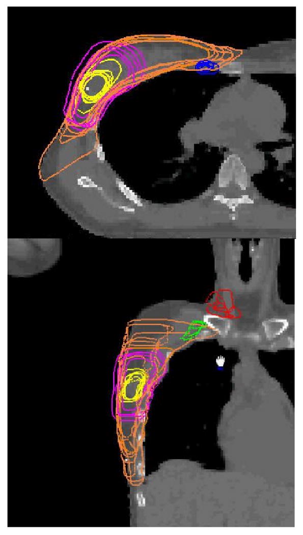
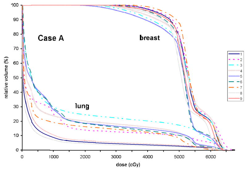
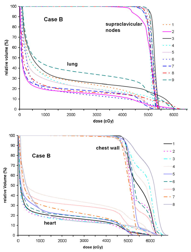
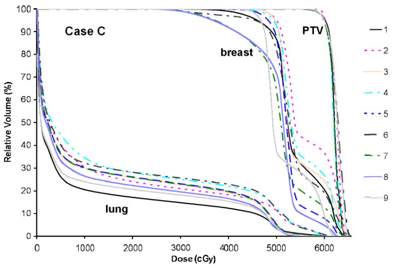
Methods and materials: Nine radiation oncologists specializing in breast RT from eight institutions independently delineated targets (e.g., lumpectomy cavity, boost planning target volume, breast, supraclavicular, axillary and internal mammary nodes, chest wall) and OARs (e.g., heart, lung) on the same CT images of three representative breast cancer patients. Interobserver differences in structure delineation were quantified regarding volume, distance between centers of mass, percent overlap, and average surface distance. Mean, median, and standard deviation for these quantities were calculated for all possible combinations. To assess the impact of these variations on treatment planning, representative dosimetric plans based on observer-specific contours were generated.
Results: Variability in contouring the targets and OARs between the institutions and observers was substantial. Structure overlaps were as low as 10%, and volume variations had standard deviations up to 60%. The large variability was related both to differences in opinion regarding target and OAR boundaries and approach to incorporation of setup uncertainty and dosimetric limitations in target delineation. These interobserver differences result in substantial variations in dosimetric planning for breast RT.
Conclusions: Differences in target and OAR delineation for breast irradiation between institutions/observers appear to be clinically and dosimetrically significant. A systematic consensus is highly desirable, particularly in the era of intensity-modulated and image-guided RT.
Conflict of interest statement
Figures
References
-
- Buchholz TA, Gurgoze E, Bice WS, et al. Dosimetric analysis of intact breast irradiation in off-axis planes. Int J Radiat Oncol Biol Phys. 1997;39:261–267. - PubMed
-
- Gray JR, McCormick B, Cox L, et al. Primary breast irradiation in large-breasted or heavy women: Analysis of cosmetic outcome. Int J Radiat Oncol Biol Phys. 1991;21:347–354. - PubMed
-
- Moody AM, Mayles WPM, Bliss JM, et al. The influence of breast size on late radiation effects and association with radiotherapy dose inhomogeneity. Radiother Oncol. 1994;33:106–112. - PubMed
-
- Taylor ME, Perez CA, Halverson KJ, et al. Factors influencing cosmetic results after conservation therapy for breast cancer. Int J Radiat Oncol Biol Phys. 1995;31:753–764. - PubMed
-
- Cheng CW, Das IJ, Baldassarre S. The effect of the number of computed tomographic slices on dose distributions and evaluation of treatment planning systems for radiation therapy of intact breast. Int J Radiat Oncol Biol Phys. 1994;30:183–195. - PubMed
Publication types
MeSH terms
Grants and funding
LinkOut - more resources
Full Text Sources
Other Literature Sources
Medical
