Review
. 2009:328:1-42.
doi: 10.1007/978-3-540-68618-7_1.

The Phycodnaviridae: the story of how tiny giants rule the world

Affiliations
Review

The Phycodnaviridae: the story of how tiny giants rule the world

W H Wilson et al. Curr Top Microbiol Immunol. 2009.

Abstract

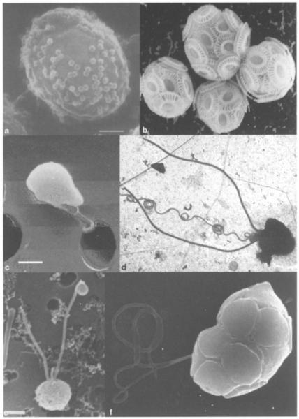
The family Phycodnaviridae encompasses a diverse and rapidly expanding collection of large icosahedral, dsDNA viruses that infect algae. These lytic and lysogenic viruses have genomes ranging from 160 to 560 kb. The family consists of six genera based initially on host range and supported by sequence comparisons. The family is monophyletic with branches for each genus, but the phycodnaviruses have evolutionary roots that connect them with several other families of large DNA viruses, referred to as the nucleocytoplasmic large DNA viruses (NCLDV). The phycodnaviruses have diverse genome structures, some with large regions of noncoding sequence and others with regions of ssDNA. The genomes of members in three genera in the Phycodnaviridae have been sequenced. The genome analyses have revealed more than 1000 unique genes, with only 14 homologous genes in common among the three genera of phycodnaviruses sequenced to date. Thus, their gene diversity far exceeds the number of so-called core genes. Not much is known about the replication of these viruses, but the consequences of these infections on phytoplankton have global affects, including influencing geochemical cycling and weather patterns.


Figures

Fig. 1
Fig. 1
Final stages of infection in the marine phytoplankton Pavlova virescens. Note the different stages of virus assembly in the cell cytoplasm. Although never characterized, these VLPs have the hallmarks of a phycodnavirus. Samples were prepared by thin-sectioning in 1978! Image was given to the author when the investigator, John Green (formerly Marine Biological Association, Plymouth), was clearing out his office prior to retirement. No information was available on the size of the scale bar
Fig. 2a-f
Fig. 2a-f
Pictures of some representative algae that are hosts for phycodnaviruses. a Scanning electron micrograph (SEM) of Chlorella NC64A with PBCV-1 particles attached to its surface (scale bar, approx. 500 run), b SEM of four Emiliania huxleyi cells (each cell, approx. 5 μm in diameter), c SEM Micromonas pusilla (scale bar, approx. 500 nm). d Transmission electron micrograph (TEM) of a Chrysochromulina sp. cell in a seawater sample taken from a Norwegian fjord (cell diameter, approx. 3 μm). e SEM of Phaeocystis sp. (strain Naples) (scale bar, approx. 1 μm). f SEM of Heterosigma akashiwo (cell diameter, approx. 12 μm). Images courtesy of (a) J.L. Van Etten (Meints et al. 1984); (b) W. Wilson (unpublished); (c) Bengt Karlson (unpublished) (d) G. Bratbak and M. Heldal (unpublished); (e) D. Vaulot (Vaulot et al. 1994); (f) S. Itakura and K. Nagasaki (unpublished).
Fig. 3
Fig. 3
Life cycle of Ectocarpus siliculosus. Zoidangium: sporangium on sporophytes, gametangium on gametophytes. Note that the sporophyte possesses two types of reproductive organs: (1) unilocular sporangia where meioses occur (R’) producing taploid (n) meiospores, and (2) plurilocular sporangia where diploid (2n) mitospores are produced by multiple mitoses. (Reproduced with permission from Müller DG et al. 1998)
Fig. 4a-d
Fig. 4a-d
Three-dimensional image reconstruction of chlorella virus PBCV-1 from cryoelectron micrographs, a The virion capsid consists of 12 pentasymmetrons (highlighted in yellow) and 20 trisymmetrons (highlighted in red and two shades of blue). A pentavalent capsomer lies at the center of each pentasymmetron. Each pentasymmetron consists of one pentamer plus 30 turners. Eleven capsorners form the edge of each trisymmetron and therefore each trisymmetron has 66 trimers. b Cross-sectional view of PBCV-1 along the twofold axis. A lipid bilayered membrane, like a railroad track, exists beneath the capsid shell (magenta arrows). Magnified views at two- and fivefold axes (outlines in b are shown in c and d, respectively), c The capsomers are interconnected by “cement” proteins (yellow arrows), d Dense material (blue arrow) (cell wall-digesting enzyme(s)?) is present at each vertex (red arrow) between the vertex and the membrane, (a reproduced with permission from Simpson et al. 2003. b-d reproduced with permission from Van Etten 2003).
Fig. 5
Fig. 5
The Phycodnaviridae capsids have 20 equilateral triangle faces composed of protein subunits and are defined by having a two-, three- and fivefold axis of symmetry. From left to right, there are six fivefold axes of symmetry passing through the vertices, ten threefold axes extending through each face and 15 twofold axes passing through the edges of an icosahedron. (Diagram produced by Thomas Locke).
Fig. 6
Fig. 6
Proposed replication cycle of chlorella virus PBCV-1. The virus uncoats at the surface of the alga and the viral DNA, possibly with associated proteins, is assumed to move to the nucleus where early gene transcription begins within 5-10 min postinfection (p.i.) The early mRNAs are transported to the cytoplasm for translation, and the early proteins presumably return to the nucleus to initiate DNA replication, which begins 60-90 min p.i., followed by late gene transcription. Late mRNAs are transported to the cytoplasm for translation, and many of these late proteins are targeted to the cytoplasmically located virus assembly centers, where virus capsids are formed. The algal cell membrane and wall lyses and infectious PBCV-1 progeny viruses are released at 6-8 h p.i. Solid arrows are known events; dotted arrows are hypothesized events. (Reproduced with permission from Kang et al. 2005).
Fig. 7
Fig. 7
Phylogenetic inference tree based on a distance matrix algorithm between concatenated conserved domains from A18-like helicase, D6R-like helicase, A32-like ATPase, DNA polymerase, thio-oxidoreductase from members of the NCLDV group (Neighbor, in PHYLIP version 3.6b). Numbers at nodes indicate bootstrap values retrieved from 100 replicates for both the neighbor-joining and parsimony analyses. The bar depicts one base substitution per ten amino acids. (Diagram adapted from Allen et al. 2006d). Viruses included are African swine fever virus (ASFV), Amsacta moorei entomopoxvirus (AMEV), Melanoplus sanguinipes entomopoxvirus (MSEV), bovine papular stomatitis virus (BPSV), fowlpox virus (FWPV), sheeppox virus (SPPX), swinepox virus (SWPX), vaccinia virus (VACV), Molluscum contagiosum virus (MOCV), myxoma virus (MYXV), Yaba monkey tumor virus (YMTV), Paramecium bursaria chlorella virus 1 (PBCV-1), Ectocarpus siliculosus virus 1 (ESV-1), Emiliania huxleyi virus 86 (EhV-86), frog virus 3 (FV3), invertebrate iridescent virus 6 (IIV6), Regina ranavirus (RRV), lymphocystis disease virus 1 (LCDV) and mimivirus.

References

    1. Agarkova IV, Dunigan DD, Van Etten JL. Virion-associated restriction endonucleases of chloroviruses. J Virol. 2006;80:8114–8123. - PMC - PubMed
    1. Allen MJ, Wilson WH. The coccolithovirus microarray: an array of uses. Brief Funct Genomic Proteomic. 2006;5:273–279. - PubMed
    1. Allen MJ, Forster T, Schroeder DC, Hall M, Roy D, Ghazal P, Wilson WH. Locus-specific gene expression pattern suggests a unique propagation strategy for a giant algal virus. J Virol. 2006a;80:7699–7705. - PMC - PubMed
    1. Allen MJ, Schroeder DC, Donkin A, Crawfurd KJ, Wilson WH. Genome comparison of two Coccolithoviruses. Virology J. 2006b;3:15. - PMC - PubMed
    1. Allen MJ, Schroeder DC, Holden MTG, Wilson WH. Evolutionary history of the Coccolithoviridae. Mol Biol Evol. 2006c;23:86–92. - PubMed

Publication types

LinkOut - more resources