Somatic, germline and sex hierarchy regulated gene expression during Drosophila metamorphosis
- PMID: 19216785
- PMCID: PMC2656526
- DOI: 10.1186/1471-2164-10-80
Somatic, germline and sex hierarchy regulated gene expression during Drosophila metamorphosis
Abstract
Background: Drosophila melanogaster undergoes a complete metamorphosis, during which time the larval male and female forms transition into sexually dimorphic, reproductive adult forms. To understand this complex morphogenetic process at a molecular-genetic level, whole genome microarray analyses were performed.
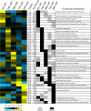
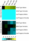
Results: The temporal gene expression patterns during metamorphosis were determined for all predicted genes, in both somatic and germline tissues of males and females separately. Temporal changes in transcript abundance for genes of known functions were found to correlate with known developmental processes that occur during metamorphosis. We find that large numbers of genes are sex-differentially expressed in both male and female germline tissues, and relatively few are sex-differentially expressed in somatic tissues. The majority of genes with somatic, sex-differential expression were found to be expressed in a stage-specific manner, suggesting that they mediate discrete developmental events. The Sex-lethal paralog, CG3056, displays somatic, male-biased expression at several time points in metamorphosis. Gene expression downstream of the somatic, sex determination genes transformer and doublesex (dsx) was examined in two-day old pupae, which allowed for the identification of genes regulated as a consequence of the sex determination hierarchy. These include the homeotic gene abdominal A, which is more highly expressed in females as compared to males, as a consequence of dsx. For most genes regulated downstream of dsx during pupal development, the mode of regulation is distinct from that observed for the well-studied direct targets of DSX, Yolk protein 1 and 2.
Conclusion: The data and analyses presented here provide a comprehensive assessment of gene expression during metamorphosis in each sex, in both somatic and germline tissues. Many of the genes that underlie critical developmental processes during metamorphosis, including sex-specific processes, have been identified. These results provide a framework for further functional studies on the regulation of sex-specific development.
Figures


Similar articles
-
Sex Differences in Drosophila Somatic Gene Expression: Variation and Regulation by doublesex.G3 (Bethesda). 2016 Jul 7;6(7):1799-808. doi: 10.1534/g3.116.027961. G3 (Bethesda). 2016. PMID: 27172187 Free PMC article.
-
Microarray analysis of Drosophila development during metamorphosis.Science. 1999 Dec 10;286(5447):2179-84. doi: 10.1126/science.286.5447.2179. Science. 1999. PMID: 10591654
-
Gene expression during the life cycle of Drosophila melanogaster.Science. 2002 Sep 27;297(5590):2270-5. doi: 10.1126/science.1072152. Science. 2002. PMID: 12351791
-
Sex and the single cell. I. On the action of major loci affecting sex determination in Drosophila melanogaster.Genetics. 1980 Feb;94(2):383-423. doi: 10.1093/genetics/94.2.383. Genetics. 1980. PMID: 6771185 Free PMC article. Review.
-
Double nexus--Doublesex is the connecting element in sex determination.Brief Funct Genomics. 2015 Nov;14(6):396-406. doi: 10.1093/bfgp/elv005. Epub 2015 Mar 22. Brief Funct Genomics. 2015. PMID: 25797692 Free PMC article. Review.
Cited by
-
Genomics of sex determination in Drosophila.Brief Funct Genomics. 2012 Sep;11(5):387-94. doi: 10.1093/bfgp/els019. Brief Funct Genomics. 2012. PMID: 23023665 Free PMC article.
-
The Wright stuff: reimagining path analysis reveals novel components of the sex determination hierarchy in Drosophila melanogaster.BMC Syst Biol. 2015 Sep 4;9:53. doi: 10.1186/s12918-015-0200-0. BMC Syst Biol. 2015. PMID: 26335107 Free PMC article.
-
Evolution of sex-specific traits through changes in HOX-dependent doublesex expression.PLoS Biol. 2011 Aug;9(8):e1001131. doi: 10.1371/journal.pbio.1001131. Epub 2011 Aug 23. PLoS Biol. 2011. PMID: 21886483 Free PMC article.
-
The ontogeny and evolution of sex-biased gene expression in Drosophila melanogaster.Mol Biol Evol. 2014 May;31(5):1206-19. doi: 10.1093/molbev/msu072. Epub 2014 Feb 12. Mol Biol Evol. 2014. PMID: 24526011 Free PMC article.
-
Everything you always wanted to know about sex ... in flies.Sex Dev. 2010;4(6):315-20. doi: 10.1159/000320632. Epub 2010 Oct 8. Sex Dev. 2010. PMID: 20926851 Free PMC article.
References
-
- Riddiford LM. Hormone receptors and the regulation of insect metamorphosis. Receptor. 1993;3:203–209. - PubMed
-
- Bodenstein D. The postembryonic development of Drosophila. In: Demerec M, editor. Biology of Drosophila. New York: Cold Spring Harbor Laboratory Press; 1950. pp. 275–367.
-
- Christiansen AE, Keisman EL, Ahmad SM, Baker BS. Sex comes in from the cold: the integration of sex and pattern. Trends Genet. 2002;18:510–516. - PubMed
-
- Manoli DS, Meissner GW, Baker BS. Blueprints for behavior: genetic specification of neural circuitry for innate behaviors. Trends in Neurosciences. 2006;29:444–451. - PubMed
Publication types
MeSH terms
Grants and funding
LinkOut - more resources
Full Text Sources
Molecular Biology Databases
Research Materials

