. 2009 Feb 24;19(4):277-86.
doi: 10.1016/j.cub.2009.01.039. Epub 2009 Feb 12.

A dynamin is required for the biogenesis of secretory organelles in Toxoplasma gondii

Affiliations

A dynamin is required for the biogenesis of secretory organelles in Toxoplasma gondii

Manuela S Breinich et al. Curr Biol. .

Abstract

Background: Apicomplexans contain only a core set of factors involved in vesicular traffic. Yet these obligate intracellular parasites evolved a set of unique secretory organelles (micronemes, rhoptries, and dense granules) that are required for invasion and modulation of the host cell. Apicomplexa replicate by budding from or within a single mother cell, and secretory organelles are synthesized de novo at the final stage of division. To date, the molecular basis for their biogenesis is unknown.

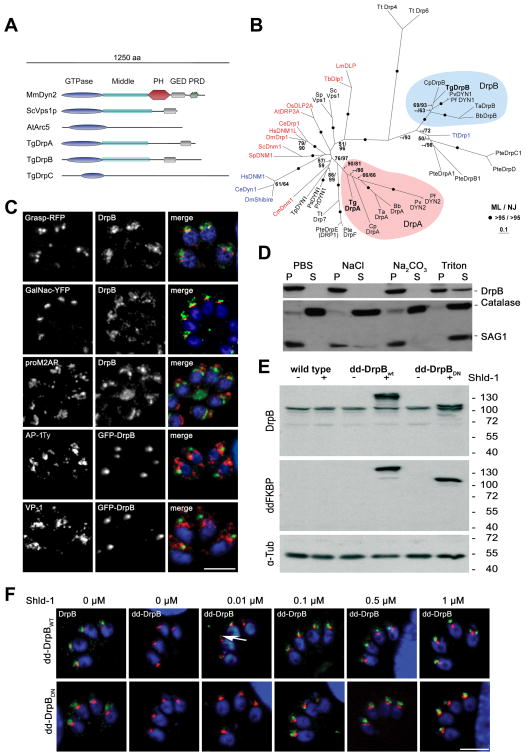
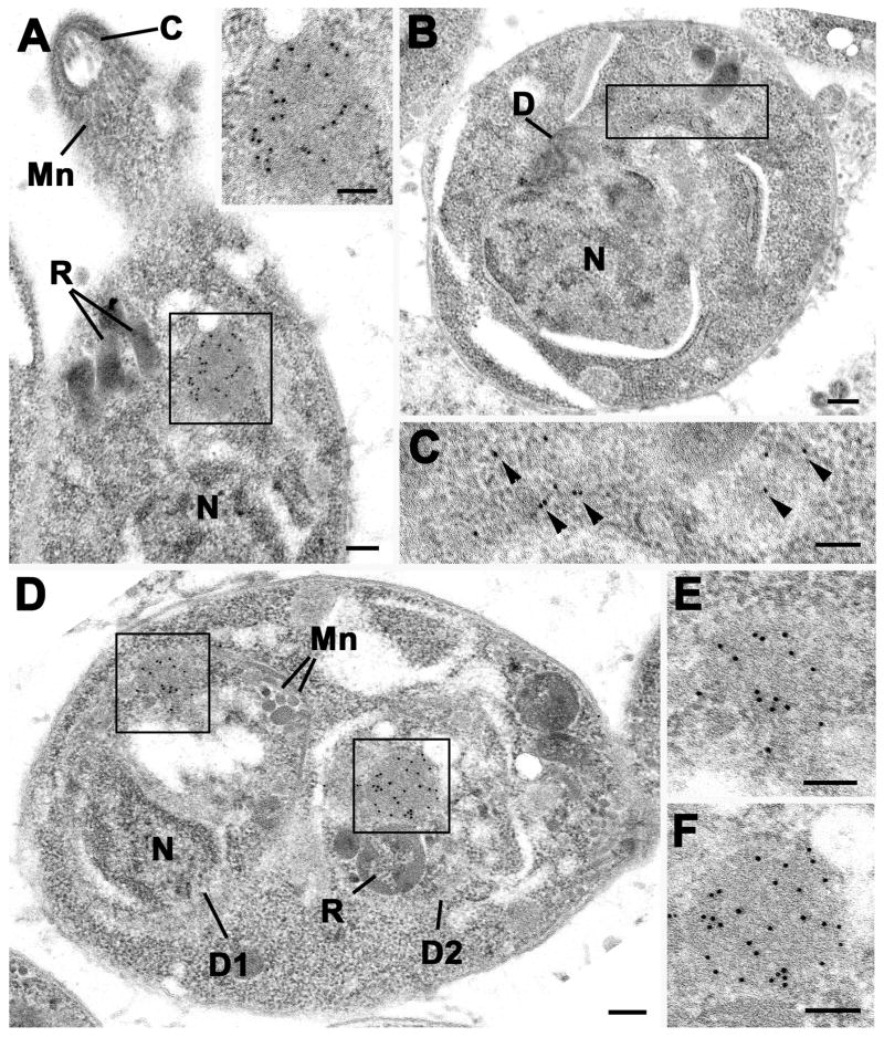
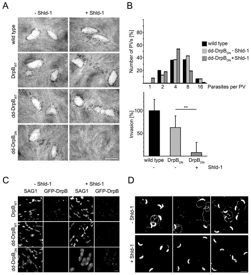
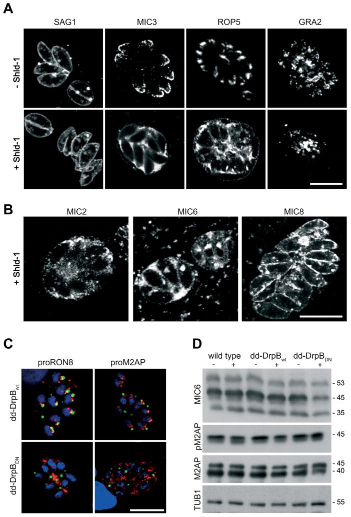
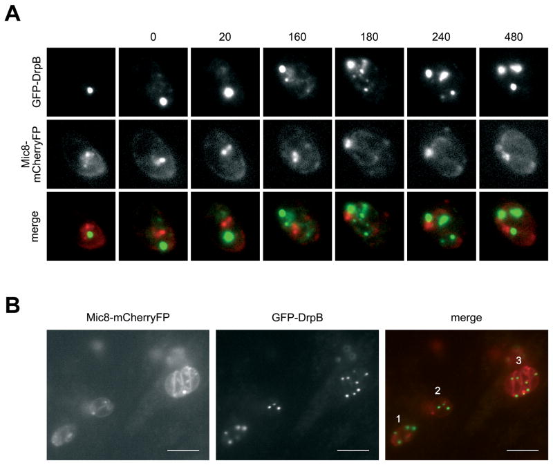
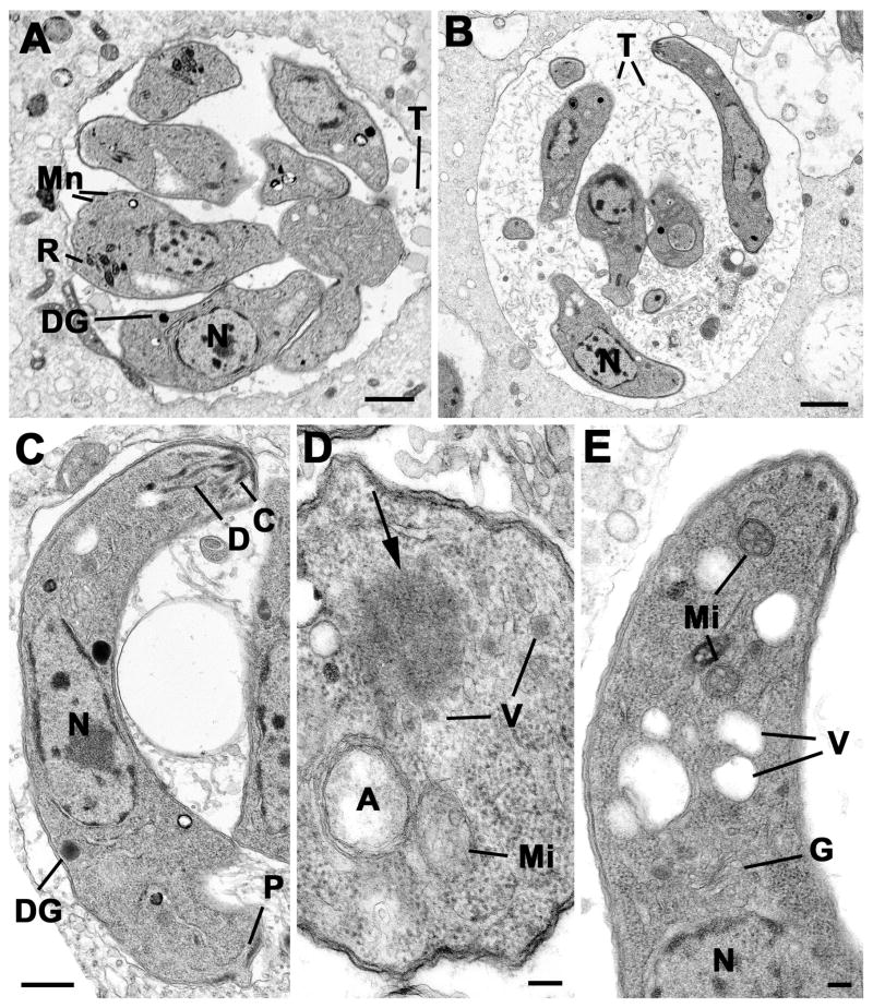
Results: We demonstrate that the apicomplexan dynamin-related protein B (DrpB) belongs to an alveolate specific family of dynamins that is expanded in ciliates. DrpB accumulates in a cytoplasmic region close to the Golgi that breaks up during replication and reforms after assembly of the daughter cells. Conditional ablation of DrpB function results in mature daughter parasites that are devoid of micronemes and rhoptries. In the absence of these organelles, invasion-related secretory proteins are mistargeted to the constitutive secretory pathway. Mutant parasites are able to replicate but are unable to escape from or invade into host cells.

Conclusions: DrpB is the essential mechanoenzyme for the biogenesis of secretory organelles in Apicomplexa. We suggest that DrpB is required during replication to generate vesicles for the regulated secretory pathway that form the unique secretory organelles. Our study supports a role of an alveolate-specific dynamin that was required for the evolution of novel, secretory organelles. In the case of Apicomplexa, these organelles further evolved to enable a parasitic lifestyle.


Figures

Figure 1
Figure 1. DrpB belongs to a ciliate specific class and localises close to the Golgi
(A) Scheme of different representative of the dynamin family, including T. gondii DrpA,B,C. Three representatives of dynamin proteins are shown considering their domain architecture (Sc: Saccharomyces cerevisiae; Mm: Mus musculus; At: Arabidopsis thaliana). (B) Phylogenetic analysis of DrpA and DrpB reveals that they belong to unrelated classes. The figure shows an unrooted Maximum Likelihood phylogenetic tree (Ln Likelihood = −33260.47) in which bootstrap values >50% based on Maximum Likelihood and Distance Matrix/Neighbor-Joining phylogenies are indicated on the respective branches. Dynamins required for endocytosis are indicated in blue and required for mitochondria division in red. DrpA and DrpB fall into two distinct clades (indicated in purple and blue respectively). The accession numbers and the alignment can be downloaded as supporting information (Bb: Babesia bovis; Ce: Caenorhabditis elegans; Cm: Cyanidioschyzon merolae; Cp: Cryptosporidium parvum; Cr: Chlamydomonas reinhardtii; Dm: Drosophila melanogaster; Hs:Homo sapiens; Lm:Leishmania major; Os:Oryza sativa; Pf:Plasmodium falciparum; Pr: Phytophthora ramorum; Ps: Phytophthora sojae; Pte: Paramecium tetraurelia; Pv: Plasmodium vivax; Sp: Schizosaccharomyces pombe; Ta: Theileria annulata; Tb: Trypanosoma brucei; Tg:Toxoplasma gondii; Tp: Thalassiosira pseudonana; Tt: Tetrahymena thermophila) (C) Immunofluorescence analysis of RHWT parasites grown on HFF-monolayers, using the indicated antibodies (proM2AP and VP-1) or transiently expressing the indicated markers (GRASP-RFP, GalNac-YFP and AP-1Ty, GFP-DrpB).. Scale bar: 10 μm (D) Cell fractionation on wild type parasites. Extracellular parasites were harvested and lysed under different conditions (PBS, PBS with 1 M NaCl, PBS with 0.1 M Na2CO3 (pH 11.5) and PBS with 2% Triton X-100) followed by ultracentrifugation. Supernatant (SN) and pellet (P) fractions were analyzed by western blot analysis with indicated antibodies. (E) Immunoblot analysis of clonal parasites transfected with dd-DrpBWT or dd-DrpBDN. Both parasite strains express the respective fusion protein only in presence of Shld-1. The immunoblot was probed with the indicated antibodies. TUB1 served as a loading control. (F) DrpBwt and DrpBDN accumulate close to the Golgi even at low overexpression. Parasites expressing GRASP-RFP and dd-DrpBwt or dd-DrpBDN were treated for 3 hours with the indicated amount of Shld-1 before localisation of GRASP and DrpB was compared. In case of dd-DrpBwt a signal for GFP can detected even at 0.01 μM Shld-1 close to the Golgi (arrow). Left: parasites not treated with Shld-1 were analysed using DrpB-antibodies. Scale bar: 10 μm. DrpB is always shown in green colour in merged images.
Figure 2
Figure 2. DrpB accumulates close to the Golgi
Electron micrographs of DrpB-GFP parasites immuno-stained with anti-GFP and visualised using 10 nm gold particles. (A) Longitudinal section through the anterior end of a tachyzoite showing a positively stained spherical structure (enclosed area) located between the nucleus (N) and rhoptries (R). Mn – microneme; C – conoid. Insert. Enlargement of the enclosed area in a showing the numerous gold particles located over the homogenous material. (B) Section through a tachyzoite at an early stage of proliferation showing early stage in formation of the daughter (D). N – nucleus. (C) Detail of the enclosed area in b showing the loss of the homogenous structure and dispersal of the gold particles (arrowheads). (D) Cross section through a tachyzoite at a late stage of division showing the two daughters (D1, D2) containing rhoptries (R) and micronemes (Mn) within the mother cell cytoplasm. (E, F) Details of the two enclose areas in D showing the reformation of the homogenous structure labelled with gold particles within the daughter organisms. Bars represent 100 nm.
Figure 3
Figure 3. Phenotypic analysis of parasites expressing DrpBDN
(A) Growth analysis of indicated parasite strains on HFF monolayers for 7 days. No plaque formation can be observed upon stabilisation of DrpBDN. Scale bar: 1 mm (B) Replication (top) and invasion (bottom) assay of indicated parasite strains. Replication was analysed after 18 hours of continuous culture in the presence or absence of Shld-1. The depicted quantification is a representative of four independent experiments. For the invasion assay parasites were grown for 36 hours in presence or absence of Shld-1. Subsequently parasites were mechanically released from their host cell and equal numbers of parasites investigated considering their ability to invade new host cell. Invasion ability of RHwt parasites was defined as 100% in each of the independent assays. Note that expression of dd-DrpBwt has no effect on invasion competency. Data represent mean values of three experiments ± s.d. Asterisks indicate significant difference (P<0.01 two-tailed Student’s t-test) (C) Egress of the host cell is blocked upon expression of DrpBDN. Indicated parasites were grown for 36 hours in presence or absence of Shld-1 and subsequently egress was triggered with A23187. After 15 minutes parasites were fixed and host cell lysis was analysed. Parasites expressing dd-DrpBDN are completely blocked in host cell egress. The depicted experiment has been repeated several times with the same result. Scale bar: 10 μm (D) Qualitative analysis of gliding motility. Parasites were treated as in b) and allowed to glide on a glass slide for 30 minutes before trail deposition was analysed with α-SAG1. Parasites expressing dd-DrpBDN are blocked in gliding. Occasionally short trails can be seen. The depicted image is a summary of three independent experiments. No difference in trail deposition was seen for RH or parasites expressing dd-DrpBWT (not shown). Scale bar: 10 μm.
Figure 4
Figure 4. DrpB is required for the transport of secretory proteins
(A, B) Immunofluorescence analysis of dd-DrpBDN-parasites grown in presence and absence of Shld-1 over night and stained with the indicated antibodies. (C) Immunofluorescence analysis of Shld-1 treated parasites expressing dd-DrpBWT and dd-DrpBDN for 24 hours in presence of Shld-1 and stained with indicated antibodies. Whereas expression of dd-DrpBWT does not interfere with the normal staining pattern of the endosomal associated compartments, in case of dd-DrpBDN expression the respective protein appears to be secreted into the lumen of the PV. Scale bar: 10 μm (D) Expression of dd-DrpBDN does not influence pro-peptide processing in intracellular parasites. For the immunoblot parasites were allowed to grow for 36 hours in presence or absence of Shld-1, before lysate was prepared and probed with the indicated antibodies. Note that a weaker signal for the mature forms of MIC6 (45kDa) and M2AP (40 kDa) were detected in Shld-1 treated dd-DrpBDN parasites (−/+ Shld-1; molecular weights are indicated). DrpB is always shown in green colour in merged images.
Figure 5
Figure 5. Time lapse analysis of parasites expressing DrpBDN
(A) Parasites co-expressing dd-DrpBDN and MIC8-mCherryFP were allowed to grow for 8 hours on HFF cells in presence of Shld-1. After this time some PVs that contain two parasites already showed mislocalisation of MIC8-mCherryFP at the plasmamembrane (see also B). In contrast single parasites show normal localisation of MIC8-mCherry in the micronemes. Image acquisition was started when re-localisation of dd-DrpBDN became apparent (t=0). One image was taken every 20 minutes. During replication strong relocalisation of dd-DrpBDN was obvious and micronemal staining of the mother cell disappeared (t=0–160). Later MIC8-mCherry gets concentrated at the posterior pole of the nascent daughter cells close but distinct to the location of dd-DrpBDN (t=180). After completion of division dd-DrpBDN accumulates in discrete spots within the two daughter cells with a significant amount also detectable at the posterior pole of the parasite (see also Fig. 3c). No staining of micronemes is detectable after replication in the mature daughter parasites (t=240–480). See also supplementary movie. (B) Endpoint of an analogous experiment. In this image three parasites are depicted. (1) parasite in the process of replication with correct localisation of MIC8-mCherry in the mother cell still detectable (note the more diffuse staining of DrpB). (2) parasites already completed one replication cycle. MIC8-mCherry shows accumulation at the posterior pole and localisation at the surface of the mature daughter parasites. In (3) the parasites completed two rounds of replication. MIC8-mCherry is found only at the surface of the parasites with no microneme staining detectable. Scale bar: 10 μm
Figure 6
Figure 6. Transmission electron micrographs of control and treated intracellular parasites
(A) Low power of a group of tachyzoites from the untreated control sample at 36 hours showing the parasites located within a relatively tight PV with few intra-vacuolar tubules (T). The tachyzoites have a central nucleus (N) and apical organelles consisting of rhoptries (R), micronemes (Mn) and dense granules (DG). Scale Bar is 1 μm. (B) Low power from at treated sample (36 hours) showing the parasites located within a loose PV with numerous intra-vacuolar tubules (T). Note the elongated appearance of the tachyzoites and the lack of apical organelles. N- nucleus. Bar is 1 μm. (C) Longitudinal section through a Shld-1 treated tachyzoites showing the centrally located nucleus (N), the conoid (C) and associated duct-like structures (D). Note the absence of rhotries and micronemes. DG – dense granule; P – posterior pore. Bar is 500 nm. (D) Cross-section through the anterior cytoplasm of a Shld-1 treated tachyzoite in which the apicoplast (A) and mitochondrion (Mi) can be seen. Note the homogenous electron dense structure (arrow) with surrounding vesicles (V). Bar is 100 nm. (E) Longitudinal section through the anterior of a Shld-1 treated tachyzoite illustrating a number of electron lucent vacuoles (V) located anterior to the Golgi body (G). N – nucleus; Mi – mitochondrion. Bar is 100 nm.

Comment in

References

    1. Carruthers V, Boothroyd JC. Pulling together: an integrated model of Toxoplasma cell invasion. Curr Opin Microbiol. 2007;10:83–89. - PubMed
    1. Sheiner L, Soldati-Favre D. Protein trafficking inside Toxoplasma gondii. Traffic. 2008;9:636–646. - PubMed
    1. Ngo HM, Yang M, Paprotka K, Pypaert M, Hoppe H, Joiner KA. AP-1 in Toxoplasma gondii mediates biogenesis of the rhoptry secretory organelle from a post-Golgi compartment. J Biol Chem. 2003;278:5343–5352. - PubMed
    1. Harper JM, Huynh MH, Coppens I, Parussini F, Moreno S, Carruthers VB. A cleavable propeptide influences Toxoplasma infection by facilitating the trafficking and secretion of the TgMIC2-M2AP invasion complex. Mol Biol Cell. 2006;17:4551–4563. - PMC - PubMed
    1. El Hajj H, Papoin J, Cerede O, Garcia-Reguet N, Soete M, Dubremetz JF, Lebrun M. Molecular signals in the trafficking of Toxoplasma gondii protein MIC3 to the micronemes. Eukaryot Cell. 2008;7:1019–1028. - PMC - PubMed

Publication types

LinkOut - more resources