. 2009 Feb 13;33(3):299-311.
doi: 10.1016/j.molcel.2009.01.019.

Fission yeast Scm3: A CENP-A receptor required for integrity of subkinetochore chromatin

Affiliations

Fission yeast Scm3: A CENP-A receptor required for integrity of subkinetochore chromatin

Alison L Pidoux et al. Mol Cell. .

Abstract

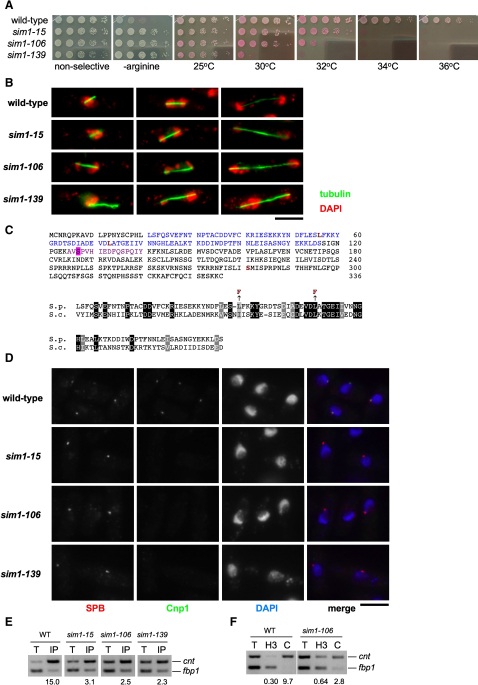
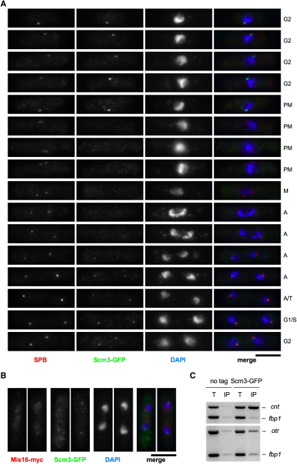
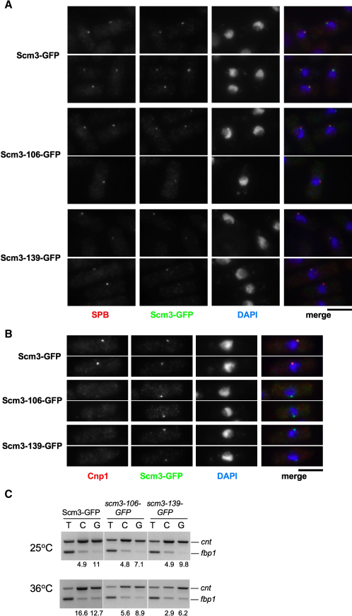
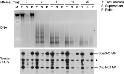
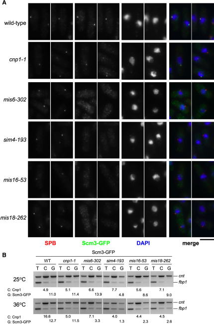
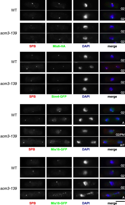
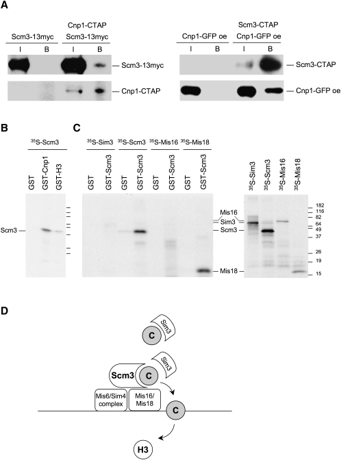
The mechanisms ensuring specific incorporation of CENP-A at centromeres are poorly understood. Mis16 and Mis18 are required for CENP-A localization at centromeres and form a complex that is conserved from fission yeast to human. Fission yeast sim1 mutants that alleviate kinetochore domain silencing are defective in Scm3(Sp), the ortholog of budding yeast Scm3(Sc). Scm3(Sp) depends on Mis16/18 for its centromere localization and like them is recruited to centromeres in late anaphase. Importantly, Scm3(Sp) coaffinity purifies with CENP-A(Cnp1) and associates with CENP-A(Cnp1) in vitro, yet localizes independently of intact CENP-A(Cnp1) chromatin and is differentially released from chromatin. While Scm3(Sc) has been proposed to form a unique hexameric nucleosome with CENP-A(Cse4) and histone H4 at budding yeast point centromeres, we favor a model in which Scm3(Sp) acts as a CENP-A(Cnp1) receptor/assembly factor, cooperating with Mis16 and Mis18 to receive CENP-A(Cnp1) from the Sim3 escort and mediate assembly of CENP-A(Cnp1) into subkinetochore chromatin.


Figures

Figure 1
Figure 1
sim1 Mutants Are Defective in Centromeric Silencing, Chromosome Segregation, and CENP-ACnp1 Association with the Centromere (A) Five-fold dilutions of the indicated strains were spotted on nonselective or medium lacking arginine to assay for alleviation of silencing at the central core (cnt1:arg3+). To assess temperature sensitivity, strains were spotted onto YES + phloxine B at the indicated temperatures; dead cells stain dark pink. (B) Chromosome segregation defects in sim1 mutants. Cells were shifted to the restrictive temperature of 36°C for 6 hr, before fixation and processing for immunofluorescence with anti-tubulin (TAT1) antibodies (green) and DAPI staining (red). Spindle length indicates that cells on left in wild-type and sim1-15 are in metaphase, and all other cells are in anaphase. Scale bar, 5 μm. (C) S. pombe Sim1 is similar to S. cerevisiae Scm3. S. pombe Sim1/Scm3 (SPAPB1A10.02) is predicted to be 37.6 kDa. Blue: equivalent to region of Scm3Sc that binds CENP-ACse4 (Mizuguchi et al., 2007). Red: amino acid changes in sim1-139 (L56F), sim1-106 (L73F), and sim1-15 (S281L). Violet: phosphorylated tryptic peptide detected by LC MS/MS at S127 (Figure S12). Bottom: Alignment of conserved region within the CENP-ACse4 binding domain of S. cerevisiae Scm3Sc (aa 74–169) with residues 21–116 of S. pombe Scm3Sp using Clustal W/BoxShade. Amino acid changes in sim1-139 (L56F) and sim1-106 (L73F) are indicated. (D) CENP-ACnp1 localization in scm3 mutants. Cells were shifted to 36°C for 6 hr, before fixation and processing for immunofluorescence with Sad1 antibodies to decorate spindle pole bodies (SPB; red) and provide a marker for approximate position of centromeres, labeled with anti-Cnp1/CENP-ACnp1 antisera (Cnp1; green). DAPI (blue). Scale bar, 5 μm. (E) ChIP for CENP-ACnp1 in the indicated strains, incubated at 36°C for 6 hr prior to fixation. Primer pairs specific for central core (cnt) and a euchromatic control locus (fbp1) were used in multiplex PCR and used to calculate enrichment in IP relative to total input chromatin (T). (F) ChIP for histone H3 (H3) and CENP-ACnp1 (C) and in wild-type and sim1-106. Primers and quantification as in (E).
Figure 2
Figure 2
Scm3Sp Is a Kinetochore Protein that Is Recruited in Late Anaphase (A) Immunofluorescence of cells expressing Scm3-GFP stained with antibodies to GFP (green) and Cdc11 (SPB; red) and DAPI (blue). Based on morphology and SPB separation, cells were assigned to cell cycle stages: G2; PM, premetaphase (prophase and prometaphase); M, metaphase; A, anaphase; T, telophase; G1; S phase. Scale bar, 5 μm. (B) Immunofluorescence of cells expressing Scm3-GFP and Mis16-myc stained with antibodies to GFP (green) and myc (red) and DAPI. Cells shown are in mid to late anaphase. (C) ChIP of Scm3-GFP using anti-GFP antibodies. Multiplex PCR indicates that Scm3-GFP is associated with central core domain (cnt) but not a euchromatic control locus (fbp1) or centromeric outer repeats (otr).
Figure 3
Figure 3
Scm3Sp Mutant Proteins Remain Localized at the Centromere, but CENP-ACnp1 Is Lost (A) Strains expressing Scm3-GFP, Scm3-106-GFP, or Scm3-139-GFP were shifted to 36°C for 6 hr, fixed, and processed for immunofluorescence using anti-GFP (Scm3-GFP; green), anti-Cdc11 antibodies (SPB; red), and DAPI (blue). Identical exposures and processing were performed, to ensure that wild-type and mutant images are comparable. All cells are in G2. Scale bar, 5 μm. (B) As (A), except antibodies are anti-GFP (green) and anti-Cnp1 (red). (C) Strains expressing Scm3-GFP, Scm3-106-GFP, and Scm3-139-GFP were shifted to 36°C for 6 hr, fixed, and analyzed by ChIP with anti-Cnp1/CENP-ACnp1 (C) and anti-GFP (G, Scm3-GFP) antibodies. PCR analysis as in Figure 1E.
Figure 4
Figure 4
Scm3Sp Does Not Extract with CENP-ACnp1 from Chromatin Extraction of CENP-ACnp1 from chromatin by MNase digestion. Nuclei were prepared from cells in which endogenous scm3+ and cnp1+ genes were TAP-tagged. After progressive MNase digestion as labeled, total nuclei digestion mixtures (T) were separated into soluble supernatant (S) and nuclei pellets (P) by centrifugation. Samples were subject to DNA extraction and electrophoresis (upper panel) or protein extraction and western blotting (lower panel). Bands labeled with an asterisk are nonspecific proteins detected by the antibody. Figure S8 shows that bulk histones (H4) are also released into supernatant.
Figure 5
Figure 5
Localization of Scm3Sp Is Dependent on Mis6, Sim4, Mis16, Mis18, but Not CENP-ACnp1 (A) Wild-type, cnp1-1, mis6-302, sim4-193, mis16-53, and mis18-262 strains expressing Scm3-GFP were shifted to 36°C for 6 hr, fixed, and processed for immunofluorescence using anti-GFP (Scm3-GFP; green), anti-Cdc11 antibodies (SPB; red), and DAPI (blue). Identical exposures and processing were performed to ensure that wild-type and mutant images are comparable. Representative images are presented. All cells are in G2. Scale bar 5 μm. (B) ChIP of Scm3-GFP in the indicated strains shifted to 36°C for 6 hr before fixation and analysis by ChIP with anti-Cnp1/CENP-ACnp1 (C) and anti-GFP (G, Scm3-GFP) antibodies. PCR analysis as in Figure 1E. Part of same experiment shown in Figure 3C; wild-type control identical.
Figure 6
Figure 6
Kinetochore Proteins Remain Localized in scm3 Mutants Wild-type and scm3-139 mutants expressing Mis6-HA, Sim4-GFP, Mis16-GFP, and Mis18-GFP were shifted to 36°C for 6 hr, fixed, and processed for immunofluorescence. For Mis6-HA cells antibodies are SPB (anti-Sad1; red) and anti-HA (Mis6-HA: green). For GFP-tagged strains antibodies are SPB (anti-Cdc11; red) and anti-GFP (Sim4, Mis16, Mis18: green). DAPI is in blue. Identical exposures and processing were performed to ensure that wild-type and mutant images are comparable. Representative images are presented. Cells were assigned to cell-cycle stages (see Figure 2A legend). Scale bar, 5 μm.
Figure 7
Figure 7
Scm3Sp Associates with Itself, CENP-ACnp1, and Mis18 (A) Coaffinity purification of Scm3Sp and CENP-ACnp1. Strains expressing the following tagged protein(s) were analyzed: Scm3-13myc only, both Scm3-13myc and CENP-ACnp1-CTAP, CENP-ACnp1-GFP only, or both CENP-ACnp1-GFP and Scm3-CTAP. Tagged genes were expressed with the native promoters at their endogenous loci, except that CENP-ACnp1-GFP was overexpressed (labeled with “oe”). IgG beads were used for affinity purification from cell extracts. Input samples (I) and bead-bound samples (B) were subject to western blotting using the indicated antibodies. (B) In vitro binding assay. (Left) 35S-labeled Scm3Sp was produced by in vitro transcription-translation in reticulocyte lysate (see [C], right panel) and incubated with GST, GST-Cnp1, or GST-H3. Complexes were pulled down with glutathione agarose, washed, and analyzed by SDS-PAGE and fluorography. (C) 35S-labeled Sim3, Scm3Sp, Mis16, and Mis18 were produced (right panel) and used in vitro binding assay with GST or GST- Scm3Sp (left panel). (D) Model for Scm3Sp function. The Mis6/Sim4 complex and Mis16/Mis18 are required to recruit Scm3Sp to centromeres. Scm3Sp acts as a receptor at the centromere for incoming CENP-ACnp1 from the Sim3 escort-chaperone. In conjunction with Mis16/Mis18 and other factors, Scm3Sp mediates the incorporation of CENP-ACnp1 (C) in subkinetochore chromatin in place of H3.

Comment in

References

    1. Ahmad K., Henikoff S. Histone H3 variants specify modes of chromatin assembly. Proc. Natl. Acad. Sci. USA. 2002;99(Suppl 4):16477–16484. - PMC - PubMed
    1. Allshire R.C., Javerzat J.P., Redhead N.J., Cranston G. Position effect variegation at fission yeast centromeres. Cell. 1994;76:157–169. - PubMed
    1. Allshire R.C., Nimmo E.R., Ekwall K., Javerzat J.P., Cranston G. Mutations derepressing silent centromeric domains in fission yeast disrupt chromosome segregation. Genes Dev. 1995;9:218–233. - PubMed
    1. Aravind L., Iyer L.M., Wu C. Domain architectures of the Scm3p protein provide insights into centromere function and evolution. Cell Cycle. 2007;6:2511–2515. - PMC - PubMed
    1. Bhargava M.M., Cramer J.H., Halvorson H.O. Isolation of high molecular weight DNA from yeast nuclei. Anal. Biochem. 1972;49:276–284. - PubMed

Publication types

MeSH terms