. 2009 Feb;16(2):271-9.
doi: 10.1016/j.devcel.2008.12.005.

Interactions with the Abelson tyrosine kinase reveal compartmentalization of eyes absent function between nucleus and cytoplasm

Affiliations

Interactions with the Abelson tyrosine kinase reveal compartmentalization of eyes absent function between nucleus and cytoplasm

Wenjun Xiong et al. Dev Cell. 2009 Feb.

Abstract

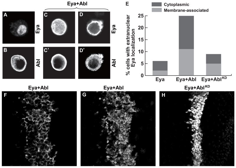
Eyes absent (Eya), named for its role in Drosophila eye development but broadly conserved in metazoa, possesses dual functions as a transcriptional coactivator and protein tyrosine phosphatase. Although Eya's transcriptional activity has been extensively characterized, the physiological requirements for its phosphatase activity remain obscure. In this study, we provide insight into Eya's participation in phosphotyrosine-mediated signaling networks by demonstrating cooperative interactions between Eya and the Abelson (Abl) tyrosine kinase during development of the Drosophila larval visual system. Mechanistically, Abl-mediated phosphorylation recruits Eya to the cytoplasm, where in vivo studies reveal a requirement for its phosphatase function. Thus, we propose a model in which, in addition to its role as a transcription factor, Eya functions as a cytoplasmic protein tyrosine phosphatase.


Figures

Figure 1
Figure 1. Genetic interactions reveal cooperativity between eya and abl
(A) Altered abl dosage dominantly modifies Eya’s ectopic eye induction efficiency. Lines 1–4 are independent transgenic lines; lof, loss-of-function; gof, gain-of-function; kd, kinase dead; n, number scored; NR, none recovered. %EE, percent of flies of genotype kinase/+; dpp-Gal4>UAS-Eya with ectopic eye tissue on head. (B) Reduced eya dosage impairs viability of abl mutant embryos. N, number of animals scored. Dissected ventral nerve cords from Stage 16 embryos stained with BP102 to reveal the pattern of the axon scaffold. (C) Wild type. (D) abl2 homozygotes have intact commissures. (E) eyaA188 homozygotes are indistinguishable from wild type. (F) eyaA188/+, abl2 mutant embryos have discontinuities along the longitudinal axon bundles with 20% of commissures lost or defective. (G) In eyaA188;abl2 double homozygotes 77% of commissures are lost and the longitudinal tracts show severe disruptions.
Figure 2
Figure 2. Eya and Abl are required for photoreceptor axon targeting in the brain
Dissected eye-brain complexes from third-instar larvae stained with anti-chaoptin 24B10 (A–D, I–K) or with anti β-galactosidase to visualize the R2–R5 specific Ro-lacZtau marker (E–H). (A,E) In wild type, R1–R6 axons form a normal lamina plexus, and R7 and R8 axons are arranged in regular staggered rows within the medulla. R2–R5 axons mostly stop in the lamina. (B,F) In abl1/abl2 trans-heterozygotes the lamina plexus is discontinuous, with gaps in the plexus, and thicker axon bundles beneath. A subset of R2–R5 axons fail to stop in the lamina and extend into the medulla. (C,G) eya2/eyaA188 mutants phenocopy abl mutants. (D,H′) eya A188/+;abl mutants have a highly disorganized lamina with thick bundles of R2–R5 axons failing to stop in the lamina. (I–K) MARCM clones of eya and abl labeled with 24B10, red. GFP, green, marks the mutant axons. (I-I″) Photoreceptor axons of large wild type clones (I inset) exhibit normal targeting to the brain. (J-J″) Axonal projections appear normal in small abl clones. Targeting of wild type axons to mutant brain tissue appears normal (J′ insets). (K-K″) Larger abl clones show fasciculation defects and laminar gaps. (L-L″) Wild type axons exhibit normal targeting to eya mutant brain tissue.
Figure 3
Figure 3. Eya and Abl interact in post-mitotic photoreceptor cells to regulate axon targeting
Dissected eye-brain complexes from third-instar larvae stained with anti β-galactosidase to visualize the Ro-lacZtau marker. (A) GMR-Gal4/+; Ro-lacZtau/+ controls show a few thin overshooting bundles. (B) UAS-EyaRNAi/+; GMR-Gal4/+; Ro-lacZtau/+ larvae have significant mistargeting defects. (C) UAS-AblRNAi/+; GMR-Gal4/+; Ro-lacZtau/+ was similar to control. (D) Double knockdown of abl and eya (UAS-EyaRNAi, UAS-AblRNAi/+; GMR-Gal4/+; Ro-lacZtau/+) causes multiple thick axon bundles to overshoot the lamina. (E) Increased Eya expression (UAS-Eya/GMR-Gal4; Ro-lacZtau/+) causes targeting defects. (F) Reduced abl expression suppresses Eya overexpression phenotypes (UAS-AblRNAi/+; UAS-Eya/GMR-Gal4; Ro-lacZtau/+). (G) Increased cytoplasmic Eya perturbs axon targeting (GMR-Gal4/+; Ro-lacZtau/UAS-Myr-EyaWT). (H) Expression of phosphatase-dead Myr-tagged Eya (GMR-Gal4/+; Ro-lacZtau/UAS-Myr-EyaK699Q) shows only mild targeting defects. (I) Reduced abl expression suppresses Myr-EyaWT phenotypes (UAS-AblRNAi/+; UAS-Myr-EyaWT/GMR-Gal4; Ro-lacZtau/+). (J) Summary of the average number of overshooting axon bundles per brain for each genotype ± standard deviation. Scoring was performed blind to the genotype. n, number of brains scored. Statistical significance (p values) were calculated using Excel’s built-in TTEST function after performing an unpaired, one-tailed T test for each pair of genotypes. All phenotypes were significant compared to control (p<.001) except that of AblRNAi (†). p-values for other relevant comparison pairs are indicated next to brackets.
Figure 4
Figure 4. Eya is a substrate of Abl
(A–D, F) Immunoblots of immunoprecipitated Flag-Eya double labeled with anti-Flag, red, and anti-phosphotyrosine (anti-pY), green. Reduced sensitivity of anti-Flag relative to anti-pY may explain the lack of complete overlap (yellow) of the two signals. (A) In transiently transfected S2 cells, Eya pY signal increases in the presence of Abl. (B) Treatment with lambda phosphatase removes the pY signal. (C) Actin-Galr4 driven coexpression of AblWT but not kinase dead AblKD, increases tyrosine phosphorylation of Flag-Eya in embryos. (D) GMR-Gal4 driven coexpression of Abl increases tyrosine phosphorylation of Flag-Eya in 3rd instar eye discs. (E) Schematic of Eya deletion constructs. (F) Abl primarily targets the PST-rich region of Eya. Lane numbering matches construct number in (E). Molecular Weight (MW) standards are indicated in Kd on left. Arrowheads on right point out IgG bands. Asterisks indicate constructs run with expected mobility in the absence of Abl. (G) In vitro kinase assay using recombinant mammalian c-Abl. Coomassie staining in left panel shows fusion protein amounts. Right panel is phosphoimager exposure of same blot showing signal in GST-EyaPST lane. (H) In vitro kinase assays using immunoprecipitated Myc-tagged Drosophila Abl from transfected S2 cells phosphorylates GST-EyaPST. Left panel, Coomassie stain; middle panel, phosphoimager exposure of same blot. Right panels show that immunoprecipitated kinase dead Abl (KD) does not phosphorylate GST-EyaPST: top, Coomassie; middle, P32 exposure; bottom, anti-Myc immunoblot; quantitation of relative intensity of signals is indicated.
Figure 5
Figure 5. Abl expression relocalizes Eya to the cytoplasm
(A–D) Indirect immunofluorescence of transfected S2 cells stained with anti-Flag to recognize Flag-Eya and/or Anti-Myc to detect Abl-Myc. (A) Eya is predominantly nuclear, although cytoplasmic staining can be seen. (B) Abl localizes to the cytoplasm with enrichment at the plasma membrane. (C–D) Cells coexpressing Eya and Abl double labeled with anti-Flag (C,D) and anti-Myc (C′,D′). (C-C′) Example of Abl expressing cell with uniform distribution of Eya. (D-D′) Example of Abl expressing cell with exclusive cytoplasmic Eya localization and membrane enrichment. (E) Quantitation of Eya localization. ~300 cells were counted for each condition. (F–H) Anti-Eya staining of wing imaginal discs coexpressing Eya and Abl transgenes with Ptc-Gal4. (F–G) Different optical sections of the same disc coexpressing Eya and Abl reveals expression of Eya in both cytoplasm (F) and nucleus (G). (H) No cytoplasmic localization was detected upon coexpression of Eya and kinase dead AblKD.
Figure 6
Figure 6. Coexpression of membrane-tethered and nuclearly-restricted Eya reveals a cytoplasmic requirement for Eya phosphatase activity
(A) Average activities of Eya transgenes in ectopic eye induction assay (see Supplementary Table 1 for details). n, number of transgenic lines tested; N, total number of flies scored. (B) Average activities of Eya transgenes in genetic rescue assay (see Supplementary Table 2 for details). n, number of transgenic lines tested; N, total number of flies scored. (C – F) Adult heads representative of: eyeless phenotype of eya2; modest rescue by NLS-EyaWT; strong rescue by coexpressed NLS-EyaWT + Myr-EyaWT; and modest rescue by coexpressed NLS-EyaWT + Myr-EyaK699Q. (G) Average ectopic eye induction efficiency of recombinant lines obtained by systematic crossing of four NLS-EyaWT lines with multiple Myr-EyaWT and Myr-EyaK699Q transgenes (see Supplementary Table 3 for details). n, number of independent recombinant lines tested; N, total number of flies scored; student t-test determined was applied to determine the p values between groups. No significant difference is determined between NLS-EyaWT and NLS-EyaWT+Myr-EyaK699Q (p=0.48).

References

    1. Abdelhak S, Kalatzis V, et al. A human homologue of the Drosophila eyes absent gene underlies branchio-oto-renal (BOR) syndrome and identifies a novel gene family. Nat Genet. 1997;15(2):157–64. - PubMed
    1. Bennett RL, Hoffmann FM. Increased levels of the Drosophila Abelson tyrosine kinase in nerves and muscles: subcellular localization and mutant phenotypes imply a role in cell-cell interactions. Development. 1992;116(4):953–66. - PubMed
    1. Bonini NM, Bui QT, et al. The Drosophila eyes absent gene directs ectopic eye formation in a pathway conserved between flies and vertebrates. Development. 1997;124(23):4819–26. - PubMed
    1. Bonini NM, Leiserson WM, et al. The eyes absent gene: genetic control of cell survival and differentiation in the developing Drosophila eye. Cell. 1993;72(3):379–95. - PubMed
    1. Bonini NM, Leiserson WM, et al. Multiple roles of the eyes absent gene in Drosophila. Dev Biol. 1998;196(1):42–57. - PubMed

Publication types