. 2009 Feb;16(2):292-302.
doi: 10.1016/j.devcel.2008.12.001.

Displacement of formins from growing barbed ends by bud14 is critical for actin cable architecture and function

Affiliations

Displacement of formins from growing barbed ends by bud14 is critical for actin cable architecture and function

Melissa Chesarone et al. Dev Cell. 2009 Feb.

Abstract

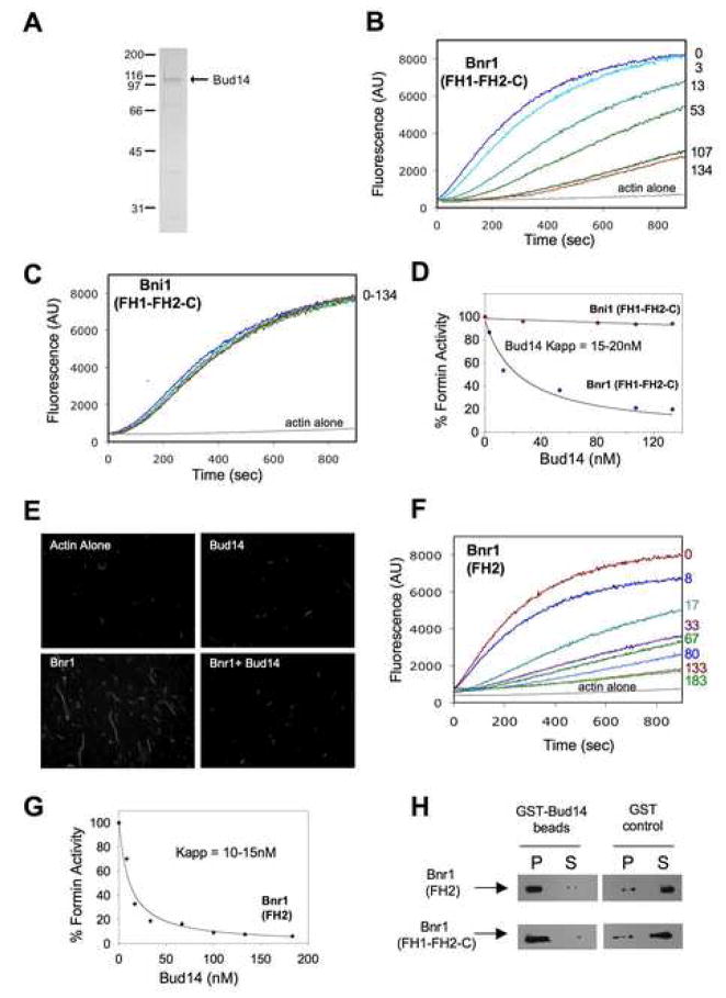
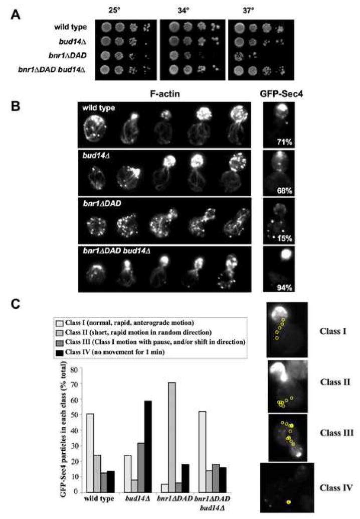
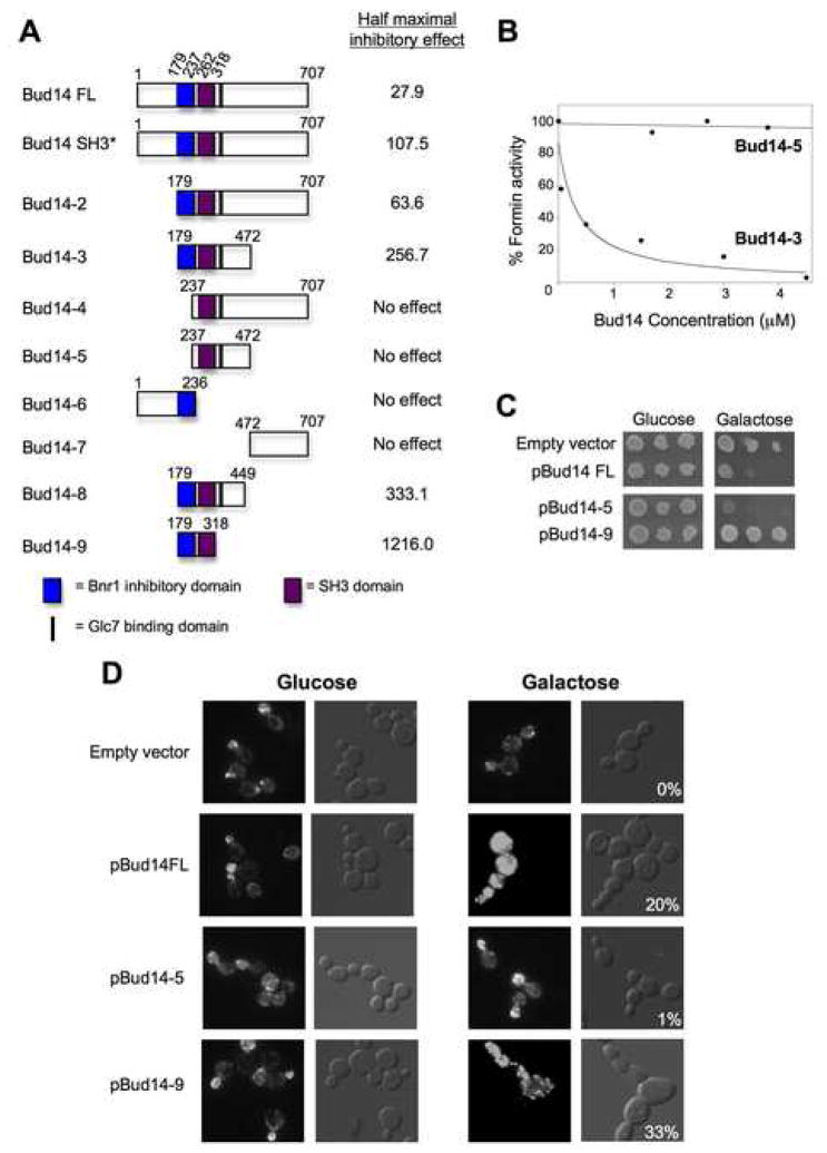
Normal cellular development and function require tight spatiotemporal control of actin assembly. Formins are potent actin assembly factors that protect the growing ends of actin filaments from capping proteins. However, it is unresolved how the duration of formin-mediated actin assembly events is controlled, whether formins are actively displaced from growing ends, and how filament length is regulated in vivo. Here, we identify Bud14 as a high-affinity inhibitor of the yeast formin Bnr1 that rapidly displaces the Bnr1 FH2 domain from growing barbed ends. Consistent with these activities, bud14Delta cells display fewer actin cables, which are aberrantly long, bent, and latrunculinA resistant, leading to defects in secretory vesicle movement. Moreover, bud14Delta suppressed mutations that cause abnormally numerous and shortened cables, restoring wild-type actin architecture. From these results, we propose that formin displacement factors regulate filament length and are required in vivo to maintain proper actin network architecture and function.


Figures

Figure 1
Figure 1. Actin cable defects in bud14Δ cells
(A) Representative architectural defects in actin cables found in bud14Δ cells. Whereas cables in wild type cells contoured the cell cortex (large arrow, upper panels), cables in bud14Δ cells were “kinked” or “buckled” (small arrow, lower panels). Cables that changed direction with > 90 degree turn at the cortex were scored as “bent”. (B) Data quantified from two experiments. (C) Total number of actin cables visible in bud14Δ versus wild type cells. Data averaged from two experiments. (D) Representative images from experiments in which wild type and bud14Δ cells were treated with 20 μM LatA for 60 seconds, fixed, and stained with Alexa-488-phalloidin to score percentage cells with visible cables. “Enhanced” panels are the same images (LatA-treated) contrast-enhanced to highlight the remaining cables. (E) Cells were treated as above with LatA for different lengths of time before fixation and imaging (0–120 sec). Data were quantified from at least two independent experiments (**=p<.001, *= p<.05). (F) Yeast strains were serially diluted and compared for growth at 25 C and 34 C on YPD plates. (G) GFP-Sec4 localization in strains grown at 25°C. Percentage of cells with a bright spot of GFP-Sec4 at the bud tip was scored (n>100), and is listed in each panel. (H) DIC microscopy of strains in F.
Figure 2
Figure 2. BUD14functions upstream ofBNR1and in parallel toBNI1
(A) Yeast strains were serially diluted and compared for growth at 25 C and 37 C on YPD plates. (B) DIC microscopy of strains grown at 25°C. (C) Percentage of cells with elongated buds was scored for each strain (n > 200) in two independent experiments.
Figure 3
Figure 3. Requirements for GFP-Bud14 localization
(A) Localization of GFP-Bud14. (B) GFP-Bud14 localization after treatment of cells with 50 μM LatA. (C) GFP-Bud14 localization after genetic disruption of actin cables or Myo2 function. Each of the indicated strains carrying GFP-Bud14 was imaged after growth at 25 C or a 10 min shift to the non-permissive temperature (37 C). (D) GFP-Bud14 localization in bnr1Δ cells. For all strains, GFP-Bud14 localization patterns were uniform in the populations.
Figure 4
Figure 4. Purified Bud14 directly inhibits Bnr1
(A) Coommassie stained gel of purified full-length Bud14. (B,C) Monomeric actin (2 μM, 5% pyrene labeled) was polymerized in the presence of (B) 5nM Bnr1(FH1-FH2-C) or (C) 30nM Bni1(FH1-FH2-C) and the indicated concentrations of Bud14. (D) Concentration-dependent effects of Bud14 on Bnr1 or Bni1 activity. Percent activity was determined by dividing the slope of the actin polymerization curve in the presence of a given concentration of Bud14 by the slope of the curve in the absence of Bud14 (considered 100% activity). (E) Visualization of filaments assembled by Bnr1 in the presence and absence of Bud14. Reaction samples were removed (same conditions as B) 10 min after initiation; filaments were stabilized with Alexa-488-phalloidin and imaged. (F) Bud14 inhibits Bnr1(FH2). Reaction conditions as in B, except using 20nM Bnr1(FH2). (G) Concentration-dependent effects of Bud14 on Bnr1(FH2) (determined as in D). (H) Association of 500nM 6His-Bnr1(FH1-FH2-C) or 6His-Bnr1(FH2) with GST-Bud14 or GST (control) immobilized on glutathione agarose. Levels of 6His-Bnr1 in the pellet (P) and supernatant (S) fractions compared by immunoblotting with 6His antibodies.
Figure 5
Figure 5. Bud14 displaces Bnr1 from growing barbed ends
(A) Effects of Bud14 on Bnr1-capped filament barbed end growth in the presence and absence of capping protein (CP). At time zero, monomeric actin (0.5μM) was added to mechanically sheared F-actin seeds in the presence of Bnr1(FH1-FH2-C), CP, and/or Bud14 (concentrations indicated in B). (B) Rates of elongation averaged from multiple experiments. (C) Elongation assays performed as in A, except instead of premixing Bnr1 with Bud14, Bud14 or control buffer was added to the reactions 65 seconds after initiation. (D) Rates of elongation averaged from multiple experiments.
Figure 6
Figure 6. bud14Δ suppresses the actin cable architecture and secretory transport defects of abnr1ΔDADstrain
(A) Yeast strains were serially diluted and compared for growth at 25°C, 34°C and 37°C on YPD plates. (B) Cells were fixed and stained with Alexa-488-phalloidin, or transformed with a GFP-Sec4 plasmid to localize secretory vesicles. Images of representative cells shown. GFP-Sec4 panels include the percentage of cells having >50% GFP-Sec4 fluorescence in the bud (n>100). (C) Time-lapse movement of GFP-Sec4 particles in live cells. The four categories of movement observed (Class I, II, III and IV) were scored in each strain (n>100 cells). Shown to the right of the bar graph are representative cells depicting each category of movement, with yellow circles tracing the path of movement recorded at 300 millisecond intervals. See supplementary materials for movies: wild type (Movie S1), bud14Δ (Movie S2), bnr1ΔDAD (Movie S3), and bud14Δbnr1ΔDAD (Movie S4).
Figure 7
Figure 7. Bud14 domain analysis
(A) Schematic of purified, truncated Bud14 polypeptides. Each Bud14 polypeptide was compared at a range of concentrations for its effects on Bnr1(FH1-FH2-C)-mediated pyrene-actin assembly. Concentrations of Bud14 required for half-maximal Bnr1 inhibition are shown. (B) Dose-dependent effects of two constructs, Bud14-5 and Bud14-3, on Bnr1(FH1-FH2-C) activity. (C) Effects on cell growth after Gal-driven overexpression of full length Bud14, Bud14-5, and Bud14-9. Cells were grown in selective media, serially diluted on selective plates containing glucose or galactose and grown at 25°C. (D) Cell morphology and actin organization defects caused by overexpression of Bud14 constructs. After 24 hr growth in galactose-containing medium, cells were fixed and stained with Alexa-488 phalloidin. DIC panels for Gal-induced cultures include percentage of cells with an obvious ‘chained’ phenotype and/or with grossly elongated or misshapen buds.

References

    1. Buttery SM, Yoshida S, Pellman D. Yeast formins Bni1 and Bnr1 utilize different modes of cortical interaction during the assembly of actin cables. Mol Biol Cell. 2007;18:1826–1838. - PMC - PubMed
    1. Cramer LP, Siebert M, Mitchison TJ. Identification of novel graded polarity actin filament bundles in locomoting heart fibroblasts: implications for the generation of motile force. J Cell Biol. 1997;136:1287–1305. - PMC - PubMed
    1. Cullen PJ, Sprague GF., Jr The Glc7p-interacting protein Bud14p attenuates polarized growth, pheromone response, and filamentous growth in Saccharomyces cerevisiae. Eukaryot Cell. 2002;1:884–894. - PMC - PubMed
    1. Evangelista M, Zigmond S, Boone C. Formins: signaling effectors for assembly and polarization of actin filaments. J Cell Sci. 2003;116:2603–2611. - PubMed
    1. Faix J, Grosse R. Staying in shape with formins. Dev Cell. 2006;10:693–706. - PubMed

Publication types