. 2009 Feb;16(2):314-20.
doi: 10.1016/j.devcel.2009.01.001.

Inactivation of murine Usp1 results in genomic instability and a Fanconi anemia phenotype

Affiliations

Inactivation of murine Usp1 results in genomic instability and a Fanconi anemia phenotype

Jung Min Kim et al. Dev Cell. 2009 Feb.

Abstract

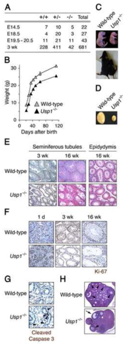
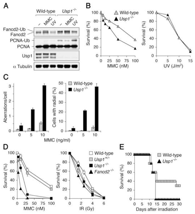
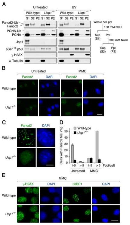
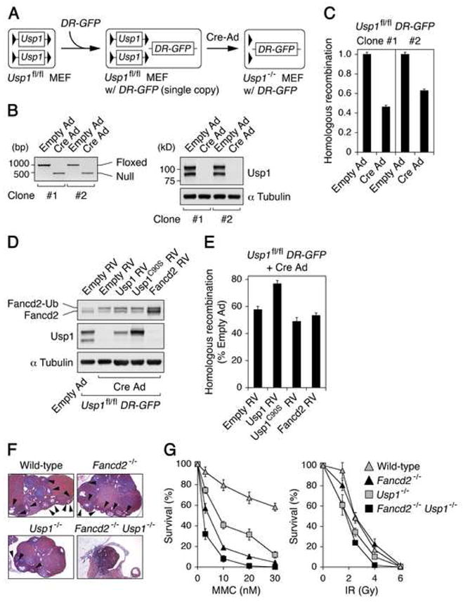
Fanconi anemia (FA) is a human genetic disease characterized by chromosome instability, cancer predisposition, and cellular hypersensitivity to DNA crosslinking agents. The FA pathway regulates the repair of DNA crosslinks. A critical step in this pathway is the monoubiquitination and deubiquitination of FANCD2. Deubiquitination of FANCD2 is mediated by the ubiquitin protease, USP1. Here, we demonstrate that targeted deletion of mouse Usp1 results in elevated perinatal lethality, male infertility, crosslinker hypersensitivity, and an FA phenotype. Usp1(-/-) mouse embryonic fibroblasts had heightened levels of monoubiquitinated Fancd2 in chromatin. Usp1(-/-) cells exhibited impaired Fancd2 foci assembly and a defect in homologous recombination repair. Double knockout of Usp1 and Fancd2 resulted in a more severe phenotype than either single knockout. Our results indicate that mouse Usp1 functions downstream in the FA pathway. Deubiquitination is a critical event required for Fancd2 nuclear foci assembly, release from chromatin, and function in DNA repair.


Figures

Figure 1
Figure 1. Perinatal lethality, growth retardation, and impaired germ cell development in Usp1-deficient mice
(A) Viability of Usp1-deficient mice at different stages of development. (B) Growth curve of wild-type (gray squares) and Usp1−/− (filled squares) mice. (C) Gross appearances of newborns and 2-week-old wild-type and Usp1−/− mice. (D) Comparison of testis size from 16-week-old wild-type and Usp1−/− mice. (E) Sections of wild-type and Usp1−/− seminiferous tubules and epididymis stained with hematoxylin-eosin. Magnification, 10 x. (F) Detection of Ki-67 by immunohistochemistry in seminiferous tubules from wild-type and Usp1−/− mice. Magnification, 40 x. (G) Detection of apoptosis by cleaved Caspase-3 staining in seminiferous tubules from 1-day-old testes. Magnification, 63 x. (H) Histological analysis of ovaries of wild-type (upper panel) and Usp1−/− (lower panel) females. Reduced number of oocytes (indicated by arrowhead) was observed in Usp1−/− ovaries, but note the presence of follicle (indicated by arrow) from which the oocyte was released during ovulation. Magnification, 5 x. For the quantitative analysis described in the text, we counted oocytes from 5 females from each genotype. We counted 4 sections per ovary (8 sections per each female; total 5 females (8X5=40 sections). All visible oocytes (including small oocytes and immediately visible oocytes) were counted.
Figure 2
Figure 2. Hypersensitivity to DNA crosslinking agents in Usp1-deficient cells
(A) Increased Fancd2-Ub and PCNA-Ub in Usp1-deficient cells. Wild-type and Usp1−/−MEFs were either left untreated, treated with MMC (500 ng/ml for 20 hr) or UV (30 J/m2, harvested at 3 hr after irradiation). Cell lysates were immunoblotted with indicated antibodies. (B) Survival rates of wild-type and Usp1−/− MEFs to MMC and UV treatment are plotted as the percentage of viable cells relative to that for respective untreated cells. (C) MMC-induced chromosomal aberrations in wild-type (open bars), Usp1+/− (gray bars) and Usp1−/− (filled bars) MEFs following the treatment with MMC for 48 hr. The numbers of chromosomal aberrations (left panel) and radial forms (right panel) per metaphase spread were scored. (D) Clonogenic survival assay of BM cells from wild-type (open squares), Usp1+/− (gray squares), Usp1−/− (filled squares) and Fancd2−/− (filled triangles) mice. BM cells were treated with increasing doses of MMC (left) or IR (right). After 7–10 days in culture, the numbers of hematopoietic colonies were compared. (E) Survival of wild-type (n = 10; gray squares) and Usp1−/− (n = 5; filled triangles) mice following 8.25 Gy of whole-body irradiation.
Figure 3
Figure 3. Increased chromatin accumulation of monoubiquitinated Fancd2 and impaired Fancd2 foci assembly in Usp1-deficient MEFs
(A) Wild-type and Usp1−/− MEFs were either left untreated or treated with UV (30 J/m2). Cells were collected 3 hr after UV irradiation and fractionated. Each fraction was immunoblotted with the indicated antibodies. (B and C) Wild-type and Usp1−/− MEFs were stained for Fancd2 following either mock treatment (untreated) or 500 ng/ml of MMC for 20 hr. Bar, 10 μm (B) and 5 μm (C). (D) Quantification of cells with Fancd2 foci. Values represent the mean ± SEM, examined at least 700 nuclei each in three independent experiments. (E) Wild-type (upper panels) and Usp1−/− (lower panels) MEFs were stained for γ-H2AX and 53BP1 foci following MMC treatment (500 ng/ml) for 20 hr. Bar, 10 μm.
Figure 4
Figure 4. HR defect in Usp1-deficient MEFs
(A) The scheme for the generation of Usp1−/− MEFs with DR-GFP reporter. (B) Confirmation of Cre-mediated excision of the loxP-flanked Usp1 gene by genomic PCR (left panel) and Western blot (right panel). (C) Two independent Usp1fl/fl DR-GFP MEF clones were analyzed for HR frequencies, each with triplicates. Values were normalized for the transfection efficiency and were displayed as mean ± SEM GFP+ frequencies relative to that of each Usp1fl/fl DR-GFP clone. (D) Immunoblotting of Usp1fl/fl DR-GFP MEFs expressing either wild-type Usp1, Usp1C90S or Fancd2 following Cre-expressing adenovirus infection. (E) I-SceI-induced HR frequencies in Usp1−/− DR-GFP MEFs transduced with empty retroviral vector or the retroviral vector encoding wild-type Usp1, Usp1C90S or Fancd2. Values were normalized for the transfection efficiency and were displayed as mean ± SEM GFP+ frequencies relative to that of each Usp1fl/fl DR-GFP MEFs. (F) Histopathology of ovaries from 12-week-old wild-type, Fancd2−/−, Usp1−/− and Usp1−/−Fancd2−/− siblings generated by Usp1+/−Fancd2+/− crosses. Usp1−/−Fancd2−/−double knockout females had a more severe ovarian atrophy than either single knockout mice. Magnification, 5 x. Arrowheads, oocytes. (G) Survival rates of BM cells from wild-type (gray triangles), Usp1−/− (gray squares), Fancd2−/− (filled triangles) and Usp1−/− Fancd2−/− (filled squares) mice following the treatment with increasing doses of MMC (left panel) or IR (right panel). After 7–10 days in culture, the numbers of hematopoietic colonies were compared.

References

    1. Agoulnik AI, Lu B, Zhu Q, Truong C, Ty MT, Arango N, Chada KK, Bishop CE. A novel gene, Pog, is necessary for primordial germ cell proliferation in the mouse and underlies the germ cell deficient mutation, gcd. Hum Mol Genet. 2002;11:3047–3053. - PubMed
    1. Chen M, Tomkins DJ, Auerbach W, McKerlie C, Youssoufian H, Liu L, Gan O, Carreau M, Auerbach A, Groves T, et al. Inactivation of Fac in mice produces inducible chromosomal instability and reduced fertility reminiscent of Fanconi anaemia. Nature Genetics. 1996;12:448–451. - PubMed
    1. Cheung AM, Elia A, Tsao MS, Done S, Wagner KU, Hennighausen L, Hakem R, Mak TW. Brca2 deficiency does not impair mammary epithelium development but promotes mammary adenocarcinoma formation in p53+/− mutant mice. Cancer Res. 2004;64:1959–1965. - PubMed
    1. Cohn MA, Kowal P, Yang K, Haas W, Huang TT, Gygi SP, D'Andrea AD. A UAF1-containing multisubunit protein complex regulates the Fanconi anemia pathway. Mol Cell. 2007;28:786–797. - PubMed
    1. Dorsman JC, Levitus M, Rockx D, Rooimans MA, Oostra AB, Haitjema A, Bakker ST, Steltenpool J, Schuler D, Mohan S, et al. Identification of the Fanconi anemia complementation group I gene, FANCI. Cell Oncol. 2007;29:211–218. - PMC - PubMed

Publication types