. 2009 May;97(1):65-74.
doi: 10.1016/j.ymgme.2009.01.006. Epub 2009 Feb 13.

Participation of asparagine 370 and glutamine 235 in the catalysis by acid beta-glucosidase: the enzyme deficient in Gaucher disease

Affiliations

Participation of asparagine 370 and glutamine 235 in the catalysis by acid beta-glucosidase: the enzyme deficient in Gaucher disease

Benjamin Liou et al. Mol Genet Metab. 2009 May.

Abstract

The hydrolysis of glucosylceramide by acid beta-glucosidase proceeds via a two-step, double displacement mechanism that includes cleavage of the O-beta-glucosidic bond, enzyme-glucosylation and, then, enzyme-deglucosylation. Two residues that may impact this cycle are N370 and E235. The N370S mutant enzyme is very common in Gaucher disease type 1 patients. Homology and crystal data predictions suggested that E235 is the acid/base catalyst in the hydrolytic reaction. Here, the roles of N370 and E235 in hydrolysis were explored using mutant proteins with selected amino acid substitutions. Heterologously expressed enzymes were characterized using inhibitors, activators, and alternative substrates to gain insight into the effects on the glucosylation (single turnover) and deglucosylation (transglucosylation) steps in catalysis. Specific substitutions at N370 selectively altered only the glucosylation step whereas N370S altered this and the deglucosylation steps. To provide functional data to support E235 as the acid/base catalyst, progress curves with poor substrates with more acidic leaving groups were used in the presence and absence of azide as an exogenous nucleophile. The restoration of E235G activity to nearly wild-type levels was achieved using azide with 2,4-dinitrophenyl-beta-glucoside as substrate. The loss of the acidic arm of the pH optimum activity curve of E235G provided additional functional support for E235 as the acid/base in catalysis. This study provides insight into the function of these residues in acid beta-glucosidase active site function.


Figures

Figure 1
Figure 1. Schematic of the double displacement mechanism of acid β-glucosidase hydrolysis of substrates
E340 and E235 are the nucleophile and putative acid/base within the active site. This retaining β-glucosidase forms a glucosyl-enzyme complex following attack of the acid of the acid/base residue (E235) at the anomeric carbon with the formation of a partially stabilized oxycarbonium structure. Ceramide (ROH) is released and a covalent glucosyl-enzyme is formed with the E340 nucleophile. The mechanism might include a carbenion rather than the partially stabilized intermediates as depicted [19]. This complex is then attacked by water that is base-assisted from the acid/base. β-glucose, the E235 and E340 are regenerated. δ designates a partial charge (− or +).
Figure 2
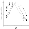
Figure 2. pH activity profiles for N370 substituted acid β-glucosidases
In (A) the TC/TX or (B) brain phosphatidylserine (32 μM)-based assays were used with the 4MU-Glc substrate. All activities were normalized to their individual maximal activities (100%) for profile comparative purposes. N370 is the wild-type enzyme. Citrate/phosphate (0.04M) buffers were used throughout. N370 (■); N370S (●); N370D (◆); N370E (▼); N370Q; (▲), D=aspartate, E=glutamate, Q=glutamine, N=asparagine, S=serine.
Figure 3
Figure 3. Activation of N370 substituted acid β-glucosidases by negatively-charged lipids and/or saposin C
In (A) Triton X-100 (4 mM) was included in each assay with taurocholate varying from 0–8 mM at pH 5.6. In (B) each assay contained brain phosphatidylserine (BPS, 0–8 μM) and the reactions were at pH 5.5. In (C) BPS and Saposin C (125 nM) were included in the assays (pH 5.5). For fold change the data were individually normalized to the respective activities without the effector(s). N370 (■); N370S (●); N370D (◆); N370E (▼); N370Q (▲).
Figure 4
Figure 4. Progress curves for the release of 2,4-dinitrophenol from the single turnover substrate, 2,4-dinitrophenyl-2-fluoro-2-deoxy-β-glucose
In (A) or (B) Triton X-100/taurocholate or BPS assay systems were used, respectively. “Active” active sites were equalized by prior titration with the DNPFG single turnover substrate/inhibitor. After about 3000–3600 sec., the absorbance from DNP plateaued at essentially equal levels for all GCases indicating an equal number of “active” active sites. Comparisons of the times to complete (2-fluoro)-glucosylation of the active sites provide relative rates of glucosylation since the Km for all the mutants were the same (108±17 μM) with the DNPG substrate. The results are composites of triplicate assays (solid lines) and their respective 95% confidence intervals (dashed lines) smoothed by non-linear one phase association using least squares from >500 data points for each curve (<3000 sec). N370S and N370E showed rates of glucosylation 10–12 fold less than those for N370 in either system. Only the N370D enzyme showed a difference in glucosylation rates with the different assay systems, indicating steric/charge changes induced by the different negatively charged lipids that specifically altered this residue’s interaction in the active site.
Figure 5
Figure 5. Transglucosylation by N370 mutant enzymes using pentanol as the acceptor
In either the TC/TX (A) or BPS-based (B) systems, N370S showed a greater fold-increase in transglucosylation with increasing pentanol than the N370 enzyme. In comparison, the other N370substituted enzymes had very similar transglucosylation enhancements by pentanol. These results are consistent with defective deglucosylation of the glucosylated N370S enzyme compared to wild-type or the other N370-substituted mutant enzymes.
Figure 6
Figure 6. Activity of E325G in the presence of azide
In (A) E325G had little activity (<0.5% WT) toward the 2,4-dinitrophenyl-β-D-glucoside substrate in the absence of azide. At 10 μM azide, the hydrolytic rate of 2,4-DNP-β-glucose by E235 (▲…. ▲) was that (95%) of the wild-type (■- ■). Except for an initial lag phase, the progress curve for generation of 2,4-dintrophenol was parallel to that of wild-type, i.e., the same DNP generation rates. In (B) azide [50 (lower) and 100 (upper) μM] were used and DNPFG was the substrate. The quantification of active sites was independent of [azide] >100 sec. The solid lines are means of two triplicate sets and the dashed lines are 95% confidence limits of the smoothed progress curves. In (C) the pH optimum curves for E325G show essentially no change in activity (<0.01 WT) in the absence of azide with DNPG as substrate (▲…. ▲). In the presence of azide the RA increased to 80–90% of wild-type with concomitant loss of the acidic arm (■--- ■). Azide had no effect on the wild-type enzyme (●-●). RA=relative activity to wild-type.

Similar articles

Cited by

References

    1. Beutler E, Grabowski GA. Gaucher disease. In: Scriver CR, Beaudet AL, Sly WS, Valle D, editors. The Metabolic and Molecular Bases of Inherited Diseases. McGraw-Hill; New York: 2001. pp. 3635–3668.
    1. Brady RO, Kanfer JN, Shapiro D. Metabolism of glucocerebrosides. II. Evidence of an enzymatic deficiency in Gaucher’s disease. Biochem Biophys Res Commun. 1965;18:221–225. - PubMed
    1. Berg-Fussman A, Grace ME, Ioannou Y, Grabowski GA. Human β-glucosidase: N-glycosylation site occupancy and the effect of glycosylation on enzymatic activity. J Biol Chem. 1993;268:14861–14866. - PubMed
    1. Grace ME, Grabowski GA. Human acid β-glucosidase: Glycosylation is required for catalytic activity. Biochem Biophys Res Commun. 1990;168:771–777. - PubMed
    1. Erickson AH, Ginns EI, Barranger JA. Biosynthesis of the lysosomal enzyme glucocerebrosidase. J Biol Chem. 1985;260:14319–14324. - PubMed

MeSH terms