. 2009 Feb 18;96(4):1307-17.
doi: 10.1016/j.bpj.2008.11.019.

Correlated motions and interactions at the onset of the DNA-induced partial unfolding of Ets-1

Affiliations

Correlated motions and interactions at the onset of the DNA-induced partial unfolding of Ets-1

Hiqmet Kamberaj et al. Biophys J. .

Abstract

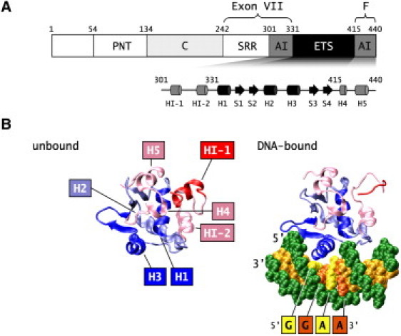
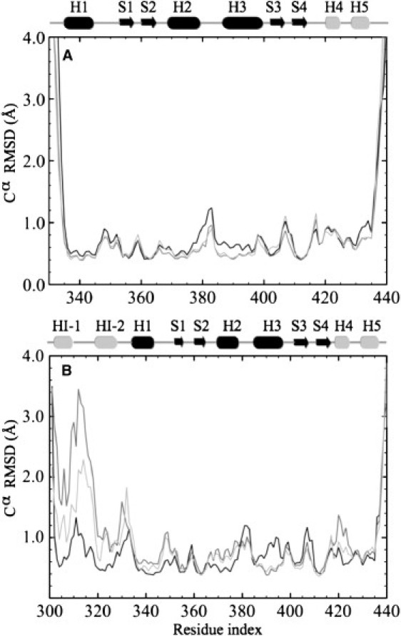
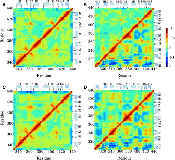
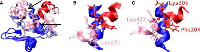
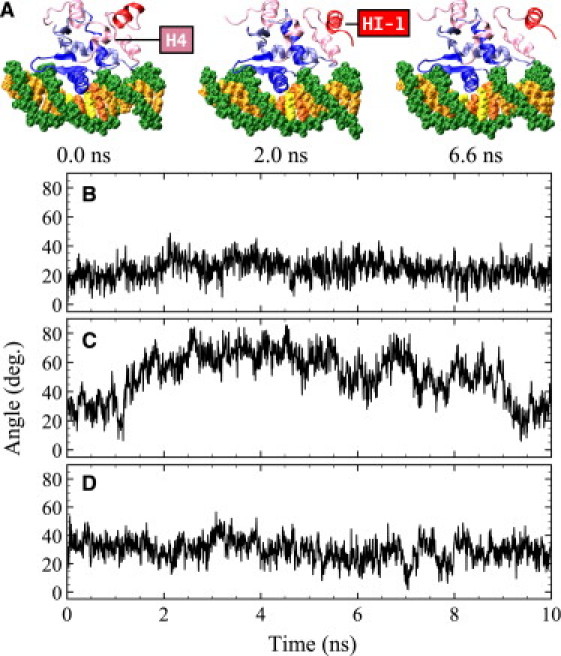
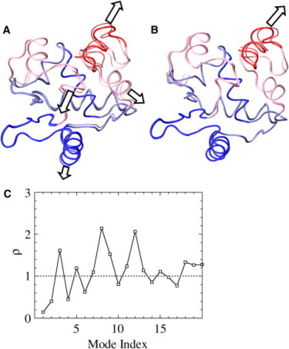
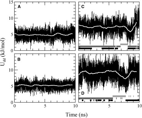
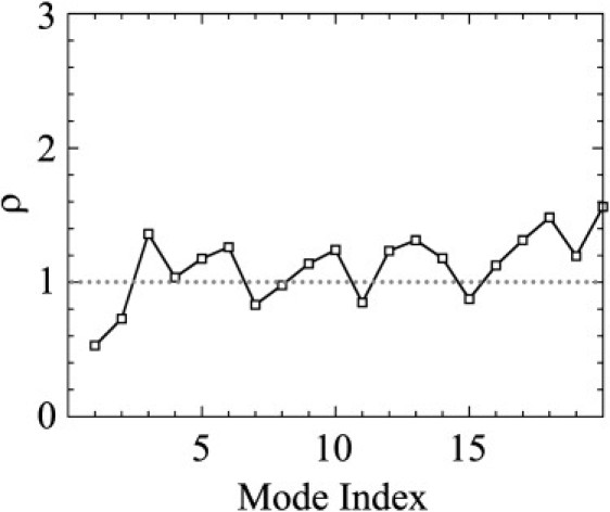
The binding of the Ets-1 transcription factor to its target DNA sequence is characterized by a highly unusual conformational change consisting of the unfolding of inhibitory helix 1 (HI-1). To probe the interactions that lead to this unfolding, we performed molecular dynamics simulations of the folded states of apo-Ets-1 and the Ets-1-DNA complex. The simulations showed large differences in correlated motions between helix 4 (H4) and HI-1. In apo-Ets-1, H4 and HI-1 moved in-phase and stabilized each other by hydrogen bonding and macrodipolar interactions, whereas in the DNA-bound state, the motion was out-of-phase, with a disruption of the stabilizing interactions. This change in motion was due to hydrogen-bonding interactions between helix 1 (H1) and the DNA. The dipolar energy between H1 and H4 was modulated by hydrogen bonds between H1 and DNA, and, in accordance with experiments, elimination of the hydrogen bonds increased the stability of HI-1. The simulations confirm that the hydrogen bonds between H1 and DNA act as a conformational switch and show that the presence of DNA is communicated from H1 to H4, destabilizing HI-1. The calculations reveal a critical role for correlated motions at the onset of the DNA-induced unfolding.


Figures

Figure 1
Figure 1
Structure of Ets-1. (A) Domain structure of Ets-1 and the secondary structure of the autoinhibitory module (AI) and the ETS domain. (B) Structure of apo-Ets-1 Δ301 (left) and the Δ301-DNA complex (right). DNA is recognized by the winged helix-turn-helix motif of the ETS domain. In apo-Ets-1, HI-1 is folded into an α-helix; upon binding the GGAA/T core sequence HI-1 unfolds. (Figs. 1B, 4, 5A, 6A and 6 B were prepared with VMD (83) and povray (www.povray.org).)
Figure 2
Figure 2
RMSD with the averaged structures for the Cα atoms of apo-Ets-1 (black) and the Ets-1-DNA complex (dark gray); the RMSD for the DNA-bound electrostatic mutant is shown in light gray. (A) Δ331. (B) Δ301.
Figure 3
Figure 3
Variance-covariance matrices of Cα fluctuations. (A and B) The upper and lower triangular parts represent the wild-type apoprotein and DNA-bound state, respectively, for Δ331 (A) and Δ301 (B). (C and D) The upper and lower triangular parts represent the electrostatic mutant and wild-type DNA-bound states, respectively, for Δ331 (C) and Δ301 (D).
Figure 4
Figure 4
Structure of apo-Δ301. (A) Overlay of the NMR and simulation structure of apo-Δ301. H5 and H4 of the NMR structure are indicated by arrows; the backbone RMSD is 2.3 Å. (B and C) H1, HI-1, and H4 of the NMR structure (B) and the simulation structure (C), with Phe304, Lys305, Leu421, and Leu422 shown as stick models. There is a hydrogen bond between Lys305 and Leu422 in both models, and an additional hydrogen bond between Phe304 and Leu422 in the simulation model. The orientation of H1 is identical in both models.
Figure 5
Figure 5
Snapshots of the simulation and angles between H4 and HI-1. (A) Structures from the MD simulations of the DNA-bound states of Δ301. Shown are the initial structure and the structure at 2 ns and 6.6 ns. (B) apo-Δ301. (C) Δ301-DNA complex. (D) Electrostatic-mutant Δ301 bound to DNA.
Figure 6
Figure 6
Quasiharmonic analysis of DNA-bound Δ301. (A) First quasiharmonic mode. The mode oscillates between the thick- and thin-lined structures; the arrows indicate major motions. (B) Second quasiharmonic mode. (C) Projection of the motion of the DNA-bound state onto the apo state of Δ301. Shown are the values of ρbound, i for the first 20 quasiharmonic modes (indexed by i). These modes account for 75% of the motion in apo-Ets-1 and for 90% of the motion in the Ets-1-DNA complex. For ρbound, i > 1, the amplitude of motion is larger in the apoprotein, whereas for ρbound, i < 1, the amplitude is larger in the DNA-bound state. The first 10 quasiharmonic modes are described in the text.
Figure 7
Figure 7
Macrodipolar interaction energy between H1 and H4, with running averages shown as white lines, for (A) apo-Δ301, (B) apo-Δ331, (C) DNA-bound Δ301, and (D) DNA-bound Δ331. In C and D, below the curves, hydrogen bonding between Gln336 and DNA is indicated by black bars, between Leu337 and DNA by dark gray bars, and no hydrogen bonding between H1 and DNA by light gray bars.
Figure 8
Figure 8
Projection of the motion of the DNA-bound state of Δ301 onto the motion of the DNA-bound state of the electrostatic mutant Δ301. Shown are the values of ρwildtype, i for the first 20 quasiharmonic modes (indexed by i). For ρwildtype, i > 1 the amplitude of motion is larger in the mutant, whereas for ρwildtype, i < 1 the amplitude is larger in the wild-type protein.

Similar articles

Cited by

References

    1. Wasylyk B., Hahn S., Giovane A. The Ets family of transcription factors. Eur. J. Biochem. 1993;211:7–18. - PubMed
    1. Maroulakou I., Bowe D. Expression and function of Ets transcription factors in mammalian development: a regulatory network. Oncogene. 2000;19:6432–6442. - PubMed
    1. Lelièvre E., Lionneton F., Soncin S., Vandenbunder B. The Ets family contains transcriptional activators and repressors involved in angiogenesis. Int. J. Biochem. Cell Biol. 2001;33:391–407. - PubMed
    1. Sato Y. Role of ETS family transcription factors in vascular development and angiogenesis. Cell Struct. Funct. 2001;26:19–24. - PubMed
    1. Oettgen P. Regulation of vascular inflammation and remodeling by ETS factors. Circ. Res. 2006;99:1159–1166. - PubMed

Publication types

LinkOut - more resources