. 2009 Feb 18;96(4):1661-70.
doi: 10.1016/j.bpj.2008.10.058.

The regulative role of neurite mechanical tension in network development

Affiliations

The regulative role of neurite mechanical tension in network development

Sarit Anava et al. Biophys J. .

Abstract

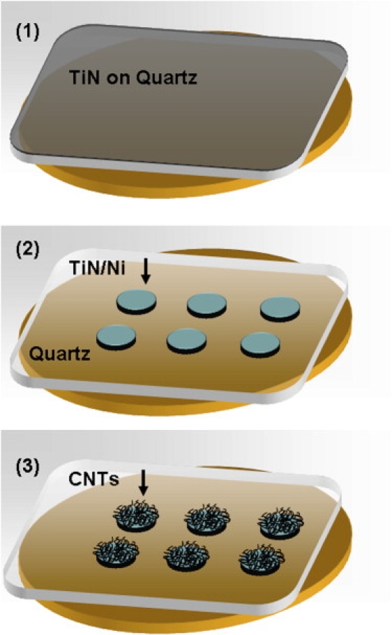
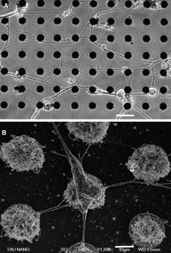
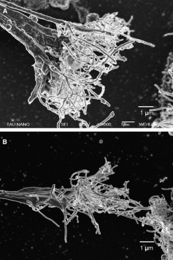
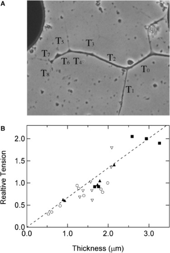
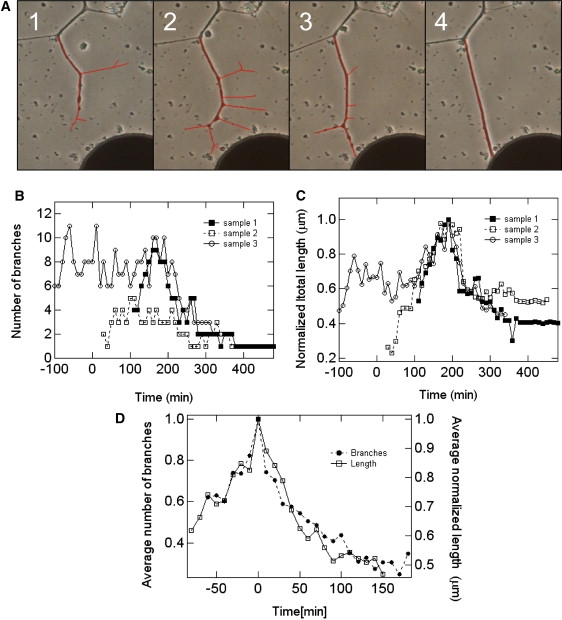
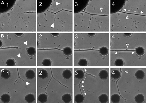
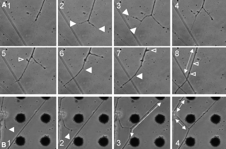
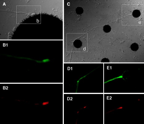
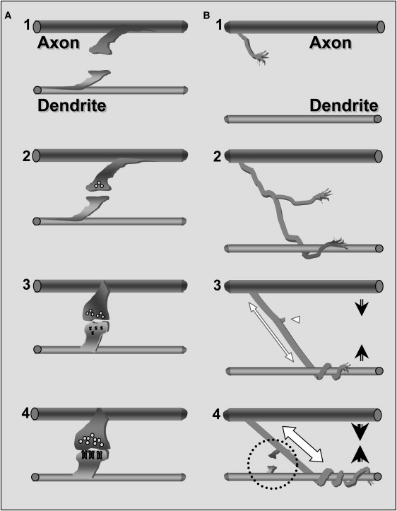
A bewildering series of dynamical processes take part in the development of the nervous system. Neuron branching dynamics, the continuous formation and elimination of neural interconnections, are instrumental in constructing distinct neuronal networks, which are the functional building blocks of the nervous system. In this study, we investigate and validate the important regulative role of mechanical tension in determining the final morphology of neuronal networks. To single out the mechanical effect, we cultured relatively large invertebrate neurons on clean quartz surfaces. Applied to these surfaces were isolated anchoring sites consisting of carbon nanotube islands to which the cells and the neurites could mechanically attach. Inspection of branching dynamics and network wiring upon development revealed an innate selection mechanism in which one axon branch wins over another. The apparent mechanism entails the build-up of mechanical tension in developing axons. The tension is maintained by the attachment of the growth cone to the substrate or, alternatively, to the neurites of a target neuron. The induced tension promotes the stabilization of one set of axon branches while causing retraction or elimination of axon collaterals. We suggest that these findings represent a crucial, early step that precedes the formation of synapses and regulates neuronal interconnections. Mechanical tension serves as a signal for survival of the axonal branch and perhaps for the subsequent formation of synapses.


Figures

Figure 1
Figure 1
Scheme of CNT microfabrication. (1) The quartz substrate with 100 nm sputtering TiN. (2) The sample after the first standard photolithography process followed by a reactive ion etching process and a second photolithography process to form a 5–7 nm of evaporated Ni catalyst layer. (3) The sample after the CVD process. The thin black lines represent the CNTs. Further details are in the text.
Figure 2
Figure 2
Culture preparation. (A) A circuit of locust neurons cultured and stabilized using 20 μm CNT islands. The cells position themselves according to the spatial distribution of the islands. The topography of the substrata also dictates the neuronal interconnections. Scale bar, 50 μm. (B) A high-resolution scanning electron microscope image of a single locust neuron adhering to an array of CNT islands; also shown are the taut neurites connecting it to neighboring islands. Scale bar, 10 μm.
Figure 3
Figure 3
Neuron attachment to the CNT. High-resolution scanning electron microscope images of the point of attachment between the terminals of neuronal processes and the CNT islands. Two examples (A and B) of the force of attachment resulting in breaking or tearing of a “handful” of tubes (shown as whitish mesh) from the CNT island, the edge of which can be seen on the right (the neurite is “arriving” from the left).
Figure 4
Figure 4
Correlation between tension and diameter of neurite segments. (A) An example of a branched axon with the series of relative tension values. Tension is approximated by measuring the angles between each angles between each two segments in every junction or brunching point. An arbitrary value is assigned T0 = 1, and the relative tension is then derived from the following relationship: Tasinθac=Tbsinθbc (as described by Bray (14))). (B) Four examples of scaled and overlaid relative tension values versus neurite segment thickness values. The scaling was carried out by multiplying all relative tension values for each of the four experiments by a constant number, so that the relative tension of the scaled data is the same for similar diameter values. The dashed line is a linear fit derived from the accumulative scaled data.
Figure 5
Figure 5
Time-lapse images (see Experimental Procedures) and analysis of process development and neurite branch pruning. (A) An extensive growth cone and neuronal arborization (1 and 2), followed by branch retraction and absorption (3 and 4). The region analyzed is traced in red. The reduction in number and total length of branches accompanies the attachment of one of the branches to a CNT island, the establishment of the connection, and the formation of tension along the axonal arbors. The course of neurite arborization described by following the change in number of secondary branches (B) or the total length of the branches (C). The independent observations were normalized by dividing the data by the maximum value. The images shown in A correspond to the data in sample 1. (D) Average curves calculated for the data shown in B (solid symbols) and C (open symbols). Data in B through D were shifted in time to align the maximum (number of branches or total length).
Figure 6
Figure 6
Three examples demonstrating the formation of tension along neurites after attachment to a CNT island (time-lapse images). The generation of tension is accompanied by the retraction and absorption of unconnected neurite branches. Solid arrowheads show newly formed branches. Open arrowheads mark the respective spots of retracted branches as tension is generated (double-headed arrows).
Figure 7
Figure 7
A process similar to the one shown in Fig. 4 is demonstrated in two examples of neurite–neurite interactions. As the connection is established, tension is generated (evidenced in the straightening of the segments), and unconnected branches are retracted and absorbed.
Figure 8
Figure 8
Neuron-CNT interactions and synaptogenesis. (A) The point of attachment of a neurite terminal to a CNT island. The area outlined and labeled as b is enlarged in B. Neuron-specific, anti-horseradish peroxidase immunostaining is shown with green in B1; antisynapsin staining is red in B2. (C) Two additional examples demonstrating established connections, evidenced by the straight segments (induced by tension). The areas outlined and marked as d and e are enlarged in D and E. The immunostaining is the same as that described and used for B.
Figure 9
Figure 9
Novel scheme of synaptogenesis. (A) The accepted stages of interneuronal connection and synapse formation (described by Cohen-Cory (3)): (1) Two-way filopodia connections; (2) an unspecialized yet functional connection is formed; (3) synaptic vesicles accumulate and postsynaptic differentiation is triggered; and (4) functional maturation of both the pre- and the postsynaptic sides. (B) The suggested preceding stages: (1) An axonal branch growth cone is approaching a dendrite; (2) entanglement of the axonal branch along the dendrite; (3) tension is formed along the axonal branch (white, double-headed arrow) as the axodendritic connection is established. The axon and dendrite are then pulled closer toward each other (arrows). The unattached branches are retracted (white arrowhead). (4) The mechanical attachment is secured as tension is increased (white, double-headed arrow). The axon and dendrite are pulled yet closer (arrows). Two-way filopodia connections are formed (dashed circle). The subsequent stages are the same as those described in A. Similar to the original model, failure at any of the stages will result in elimination of the connection.

References

    1. Luo L., O'Leary D.D. Axon retraction and degeneration in development and disease. Annu. Rev. Neurosci. 2005;28:127–156. - PubMed
    1. Hua J.Y., Smear M.C., Baier H., Smith S.J. Regulation of axon growth in vivo by activity-based competition. Nature. 2005;434:1022–1026. - PubMed
    1. Cohen-Cory S. The developing synapse: construction and modulation of synaptic structures and circuits. Science. 2002;298:770–776. - PubMed
    1. Li Z., Sheng M. Some assembly required: the development of neuronal synapses. Nat. Rev. Mol. Cell Biol. 2003;4:833–841. - PubMed
    1. Waites C.L., Craig A.M., Garner C.C. Mechanisms of vertebrate synaptogenesis. Annu. Rev. Neurosci. 2005;28:251–274. - PubMed

Publication types

Substances

LinkOut - more resources