. 2009 May;166(2):133-43.
doi: 10.1016/j.jsb.2009.02.001. Epub 2009 Feb 13.

Transient amorphous calcium phosphate in forming enamel

Affiliations

Transient amorphous calcium phosphate in forming enamel

Elia Beniash et al. J Struct Biol. 2009 May.

Erratum in

  • J Struct Biol. 2009 Jul;167(1):95

Abstract

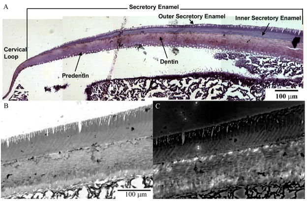
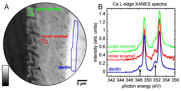
Enamel, the hardest tissue in the body, begins as a three-dimensional network of nanometer size mineral particles, suspended in a protein gel. This mineral network serves as a template for mature enamel formation. To further understand the mechanisms of enamel formation we characterized the forming enamel mineral at an early secretory stage using X-ray absorption near-edge structure (XANES) spectromicroscopy, transmission electron microscopy (TEM), FTIR microspectroscopy and polarized light microscopy. We show that the newly formed enamel mineral is amorphous calcium phosphate (ACP), which eventually transforms into apatitic crystals. Interestingly, the size, shape and spatial organization of these amorphous mineral particles and older crystals are essentially the same, indicating that the mineral morphology and organization in enamel is determined prior to its crystallization. Mineralization via transient amorphous phases has been previously reported in chiton teeth, mollusk shells, echinoderm spicules and spines, and recent reports strongly suggest the presence of transient amorphous mineral in forming vertebrate bones. The present finding of transient ACP in murine tooth enamel suggests that this strategy might be universal.


Figures

Figure 1
Figure 1
Light micrographs of semi-thin section of mouse incisor in the region of cervical loop stained with toluidine blue (A). Note differences in the shades of staining in the outer and inner sectretory enamel. Micrographs in the bottom row are taken from the same area in bright field (B) and polarized light (C) modes. Note that the inner enamel layer is slightly birefrigent.
Figure 2
Figure 2
TEM micrographs and corresponding diffraction patterns taken from unstained thin sections through outer enamel layer (A); inner enamel layer (B) and mantle dentin (C). The circle appearing in A is due to a saturation artifact, as explained in the Supplement Figure 1, and not to nanoparticulate crystals.
Figure 3
Figure 3
(A) Ca distribution map in a region of forming mouse incisor at a distance of approximately 1 mm from the cervical loop. High Ca concentration is represented by lighter gray level. The Ca map was obtained by digital ratio of 349.3 eV and 344 eV X-PEEM images. (B) XANES spectra extracted from the outer secretory enamel, inner secretory enamel and mantle dentin regions outlined in A, and correspondingly colored. Notice the significant differences in the spectral region usually affected by the crystal field (arrows).
Figure 4
Figure 4
XANES spectra acquired by X-PEEM at the Ca L-edge from the reference standard minerals (A) octacalcium phosphate (OCP), (B) hydroxyapatite (HA) (C) low crystallinity carbonated hydroxyapatite (LCHA) and (D) amorphous calcium phosphate (ACP); XANES spectra from the three regions outlined in Figures 3 and 5 are also reported: (E) inner enamel, (F) outer enamel (J) dentin and (H) outer enamel 1 year later. The best fits (gray solid line) of the experimental data (black dots) were obtained using 5 Gaussians, 2 arctangents (purple), and a third order polynomial (green). Polynomials and residues (yellow) are displaced down, data and fits are displaced up for clarity. Peaks are hereafter referred to by numbers, as shown in the HA plot. Peak 1 is the magenta Gaussian at 352.6 eV, peak 2 is the green Gaussian at 351.6 eV, etc. Notice the small dip between peaks 1 and 2, and the small separation between peaks 3 and 4 (blue and light blue) in the most amorphous phases. See Table 1 for the accurate positions of all peaks, and the crystallinity of the minerals and biominerals analyzed.
Figure 5
Figure 5
X-PEEM and TEM analysis of the outer enamel collected after 1 year ex vivo. (A) The Ca distribution map of the same area presented in the Fig. 3A. (B) A peak-fitted XANES spectrum from the region in the outer enamel outlined in green. (C) TEM micrograph of the outer secretory enamel in a 1-year-old thin section of a mouse incisor, and diffraction pattern taken from the area in the middle of the micrograph (inset). Figure 6. Second derivatives of transmission FTIR spectra obtained from the outer (A); central (B) and inner (C) early secretory enamel.
Figure 6
Figure 6
FTIR spectra taken from the outer (A), mid (B) and inner (C) layers of the early secretory enamel and their corresponding second derivative spectra (D–F).
Figure 7
Figure 7
Intermediate (A) and high magnification (B) TEM micrographs of secretory enamel at the dentin-enamel junction, collected from a fresh section. Note the chain-like texture of enamel particles in B.

Similar articles

Cited by

References

    1. Addadi L, Weiner S. Interactions Between Acidic Proteins and Crystals - Stereochemical Requirements in Biomineralization. Proc Natl Acad Sci U S A. 1985;82:4110–4114. - PMC - PubMed
    1. Addadi L, Weiner S. Control and Design Principles in Biological Mineralization. Angew Chem Int Ed Engl. 1992;31:153–169.
    1. Addadi L, Beniash E, Weiner S. Assembly and Mineralization Processes in Biomineralization: Strategies for Forming Biological Composite Materials. In: Jones W, Rao CNR, editors. Supramolecular Organization and Materials Design. Cambridge University Press; Cambridge: 2002. pp. 1–34.
    1. Aizenberg J, Muller DA, Grazul JL, Hamann DR. Direct fabrication of large micropatterned single crystals. Science. 2003;299:1205–1208. - PubMed
    1. Aoba T, Moreno E. Changes in the nature and composition of enamel mineral during procine amelogenesis. Calcif Tissue Int. 1990;47:356–364. - PubMed

Publication types

MeSH terms

Substances

LinkOut - more resources