. 2009 Feb 19;5(2):137-50.
doi: 10.1016/j.chom.2008.12.010.

PGLYRP-2 and Nod2 are both required for peptidoglycan-induced arthritis and local inflammation

Affiliations

PGLYRP-2 and Nod2 are both required for peptidoglycan-induced arthritis and local inflammation

Sukumar Saha et al. Cell Host Microbe. .

Abstract

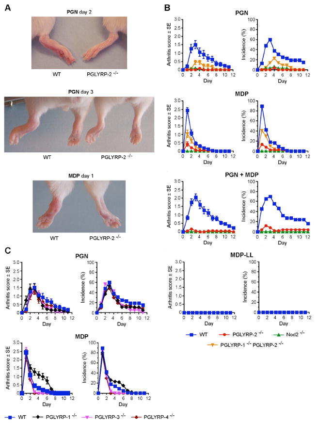
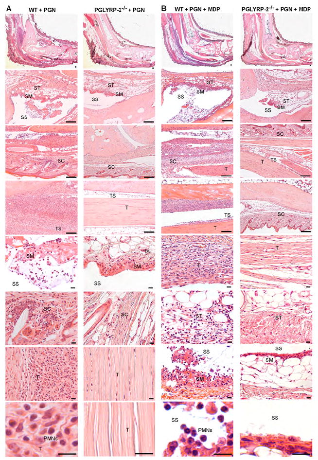
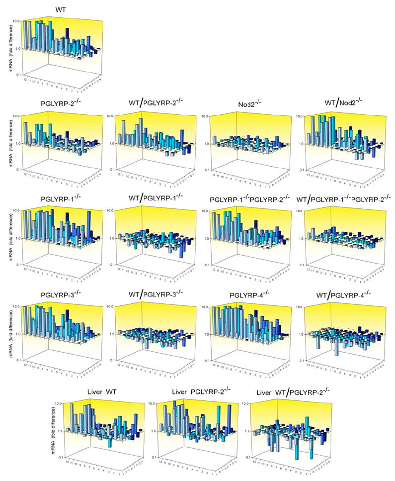
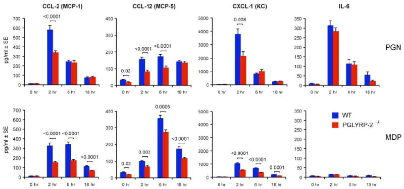
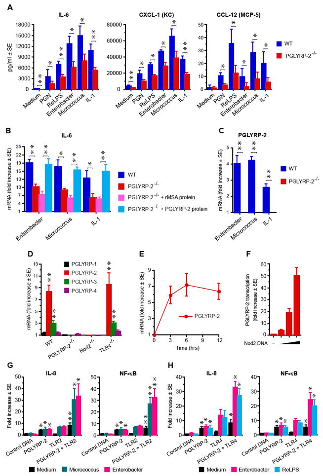
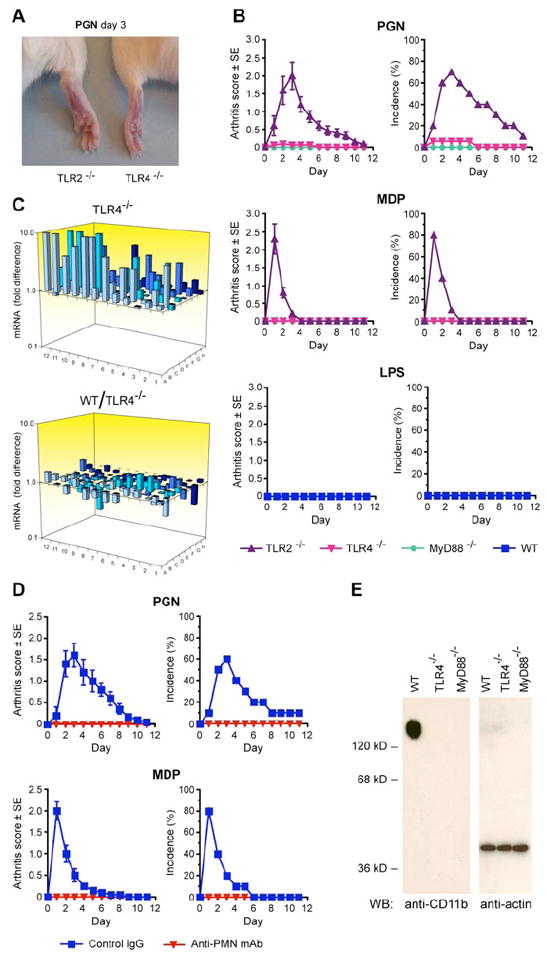
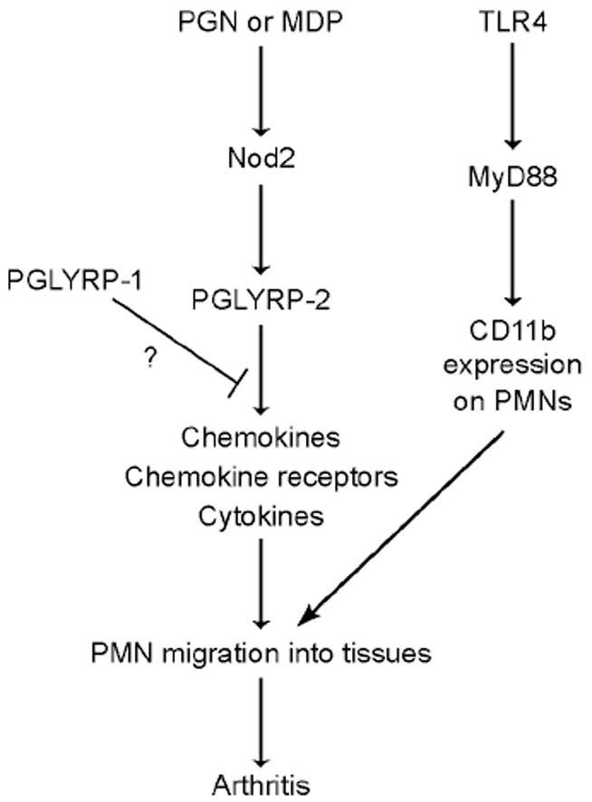
Peptidoglycan recognition proteins (PGRPs) are structurally conserved from insects to mammals. Insect PGRPs have diverse host-defense functions. Mammalian PGRPs PGLYRP-1, PGLYRP-3, and PGLYRP-4 have bactericidal activity, while PGLYRP-2 has amidase activity. To extend the known functions of mammalian PGRPs, we examined whether they have immunomodulating activities in peptidoglycan-induced arthritis in mice. We demonstrate that PGLYRP-2 and Nod2 are both required for arthritis in this model. The sequence of events in peptidoglycan-induced arthritis is activation of Nod2, local expression of PGLYRP-2, chemokine production, and recruitment of neutrophils into the limbs, which induces acute arthritis. Only PGLYRP-2 among the four mammalian PGRPs displays this proinflammatory function, and PGLYRP-1 is anti-inflammatory. Toll-like receptor 4 (TLR4) and MyD88 are required for maturation of neutrophils before peptidoglycan challenge. Our results demonstrate that PGRPs, Nod2, and TLR4, representing three different types of pattern-recognition molecules, play interdependent in vivo roles in local inflammation.


Figures

Figure 1
Figure 1. PGLYRP-2 and Nod2 are required for the development of peptidoglycan- or MDP-induced acute arthritis
(A) Swelling and erythema of ankles and feet in WT, but not in PGLYRP-2-/- mice after intravenous injection of 200 μg of peptidoglycan (PGN) or 100 μg of MDP. (B) and (C) Arthritis scores and incidence in WT or KO mice after intravenous injection of 200 μg of PGN, 100 μg of MDP, or both PGN and MDP; means ± SE of 19-76 mice/group. Arthritis scores and incidence were significantly higher in: PGN-injected, MDP-injected, and PGN+MDP-injected WT than PGLYRP-2-/- mice and Nod2-/- mice; in PGN-injected and MDP-injected WT than PGLYRP-1-/-PGLYRP-2-/- mice; and also in PGN-injected and MDP-injected PGLYRP-1-/-PGLYRP-2-/- than PGLYRP-2-/- mice (B). Arthritis scores and incidence were not significantly different in PGN-injected or MDP-injected WT than PGLYRP-1-/-, PGLYRP-3-/-, and PGLYRP-4-/- mice, except for PGLYRP-1-/- mice, which after MDP injection had significantly higher scores than WT mice (C). The P values for the significance of all the above differences are listed in Supplemental Data.
Figure 2
Figure 2. PGLYRP-2 is required for the development of peptidoglycan- or peptidoglycan+MDP-induced pathologic changes characteristic of acute arthritis
Edema and cellular infiltrations of tendons (T), tendon sheaths (TS), synovial membrane (SM), subsynovial tissue (ST), synovial space (SS), and connective and subcutaneous tissues (SC) in ankles and feet in WT, but not in PGLYRP-2-/- mice, 3 days after intravenous injection of 200 μg of PGN (A) or 200 μg of PGN + 100 μg of MDP (B). PMNs were the predominant infiltrating cells in WT mice (bottom left panels in A and B). Ankles and feet in injected PGLYRP-2-/- mice appeared normal. Bar = 100 μm in four top panels and 10 μm in four bottom panels.
Figure 3
Figure 3. MDP strongly induces chemokine and cytokine mRNA in the feet in WT, PGLYRP-1-/-, PGLYRP-3-/-, and PGLYRP-4-/-, but not in PGLYRP-2-/- and Nod2-/- mice
WT or KO mice were injected intravenously with 100 μg MDP or buffer, and after 6 hrs the amounts of mRNA for 84 pro-inflammatory genes and 5 housekeeping genes were measured in feet (or livers, bottom panels) by qPCR and expressed as the ratios of the amount of mRNA in MDP-injected to buffer-injected mice (which represents fold gene induction by MDP), or as the ratios of fold MDP-induced gene activation in WT to KO mice (which represents fold difference in the response to MDP in WT versus KO mice, WT/KO). The results are means of 12 WT or 6 KO mice/group (12 or 6 separate qPCR assays). The means, SE, the significance of differences, and the names of the genes are given in Supplemental Tables S1-3.
Figure 4
Figure 4. MDP and peptidoglycan induce higher chemokine levels in blood in WT than in PGLYRP-2-/- mice
WT or PGLYRP-2-/- mice were injected intravenously with 200 μg PGN or 100 μg MDP, and the concentrations of CCL-2, CCL-12, CXCL-1, and IL-6 were measured in the serum at the indicated times; means of 20-30 mice/group for each time point; the significance of differences between WT and PGLYRP-2-/- mice is indicated.
Figure 5
Figure 5. PGLYRP-2-/- fibroblasts have low responsiveness to bacteria and cytokines, which is reconstituted by PGLYRP-2; MDP induces expression of PGLYRP-2 in the feet through activation of Nod2; and PGLYRP-2 activates IL-8 and NF-κB and synergizes with TLR2 and TLR4
(A) IL-6, CXCL-1, and CCL-12 production by paw fibroblasts from WT and PGLYRP-2-/- mice cultured with the indicated stimulants. (B) IL-6 mRNA in paw fibroblasts from WT and PGLYRP-2-/- mice cultured with the indicated stimulants and reconstitution of the WT-level response in fibroblasts from PGLYRP-2-/- mice cultured with 1 μg/ml of recombinant mouse PGLYRP-2 protein, but not recombinant mouse albumin (rMSA). (C) PGLYRP-2 mRNA in paw fibroblasts from WT and PGLYRP-2-/- mice cultured with the indicated stimulants. (A-C) means ± SE of 3 experiments; significance of differences (WT vs PGLYRP-2-/- cells or rMSA vs PGLYRP-2 protein) is indicated by asterisks. (D) Increase in the amounts of mRNA for PGLYRP-1, PGLYRP-2, PGLYRP-3, and PGLYRP-4 in the feet of WT, PGLYRP-2-/-, Nod2-/-, and TLR4-/- mice 6 hrs after intravenous injection of 100 μg MDP, measured by qPCR and expressed as the ratios of the amount of mRNA in MDP-injected to buffer-injected mice (means ± SE of 6-12 mice/group, significance of increase is indicated by asterisks). (E) Kinetics of induction of PGLYRP-2 mRNA in the feet of WT mice after intravenous injection of 100 μg MDP (means ± SE of 6 mice/group). (F) Induction of PGLYRP-2 transcription in HEK293 cells transfected with PGLYRP-2 promoter-luciferase and control plasmid DNA (-) or increasing concentrations of Nod2 (means ± SE of 4 cultures from 2 experiments). (G, H) Induction of IL-8 transcription or NF-κB in HEK293 cells transfected with control DNA, PGLYRP-2, and TLR2 (G), or control DNA, PGLYRP-2, and TLR4 (plus CD14 and MD-2) (H), alone or in combination, after stimulation with Micrococcus, Enterobacter, or ReLPS, or no stimulation; means ± SE of 7 (G) or 4 (H) experiments, significance of differences of PGLYRP-2 vs control DNA or PGLYRP-2+TLR vs TLR is indicated by asterisks. *, P≤0.05; * *, P≤0.005.
Figure 6
Figure 6. TLR4, MyD88, and PMNs are required for the development of peptidoglycan- or MDP-induced acute arthritis
(A) Swelling and erythema of ankles and feet in TLR2-/- but not in TLR4-/- mice after intravenous injection of 200 μg of PGN. (B) Arthritis scores and incidence in TLR2-/-, TLR4-/-, and MyD88-/- mice after intravenous injection of 200 μg of PGN or 100 μg of MDP; or in WT mice after intravenous injection of 10 μg of ReLPS; means ± SE of 12-20 mice/group. (C) Expression of chemokine and cytokine mRNA in the feet of TLR4-/- mice after intravenous injection of 100 μg of MDP, determined as in Figure 3. (D) Arthritis scores and incidence in WT mice treated with control IgG or anti-PMN mAb after intravenous injection of 200 μg of PGN or 100 μg of MDP; means ± SE of 10 mice/group. (E) Blood PMNs from untreated TLR4-/- and MyD88-/- mice, in contrast to WT mice, do not express CD11b integrin detected on Western blot.
Figure 7
Figure 7. Proposed sequence of events leading to PGN- or MDP-induced local inflammation and arthritis
The chart indicates temporal sequence of events and does not imply direct signal transduction pathways. TLR4 permits maturation of PMNs before exposure to PGN or MDP.

Comment in

  • Mammalian PGRPs in the spotlight.
    Boneca IG. Boneca IG. Cell Host Microbe. 2009 Feb 19;5(2):109-11. doi: 10.1016/j.chom.2009.01.007. Cell Host Microbe. 2009. PMID: 19218081 Review.

Similar articles

Cited by

References

    1. Biragyn A, Ruffini PA, Leifer CA, Klyushnenkova E, Shakhov A, Chertov O, Shirakawa AK, Farber JM, Segal DM, Oppenheim JJ, et al. Toll-like receptor 4-dependent activation of dendritic cells by β-defensin 2. Science. 2002;298:1025–1029. - PubMed
    1. Bischoff V, Vignal C, Duvic B, Boneca IG, Hoffmann JA, Royet J. Down regulation of the Drosophila immune response by peptidoglycan-recognition proteins SC1 and SC2. PLoS Pathog. 2006;2:e14. - PMC - PubMed
    1. Chamaillard M, Hashimoto M, Horie Y, Masumoto J, Qiu S, Saab L, Ogura Y, Kawasaki A, Fukase K, Kusumoto S, et al. An essential role for NOD1 in host recognition of bacterial peptidoglycan containing diaminopimelic acid. Nat Immunol. 2003;4:702–707. - PubMed
    1. Chang YH, Pearson CM, Chedid L. Adjuvant polyarthritis. V. Induction by N-acetylmuramyl-L-alanyl-D-isoglutamine, the smallest peptide subunit of bacterial peptidoglycan. J Exp Med. 1981;153:1021–1026. - PMC - PubMed
    1. Cromartie WJ, Craddock JG, Schwab JH, Anderle SK, Yang CH. Arthritis in rats after systemic injection of streptococcal cells or cell walls. J Exp Med. 1977;146:1585–1602. - PMC - PubMed

Publication types