. 2009 May 1;284(18):12080-90.
doi: 10.1074/jbc.M807637200. Epub 2009 Feb 13.

14-3-3:Shc scaffolds integrate phosphoserine and phosphotyrosine signaling to regulate phosphatidylinositol 3-kinase activation and cell survival

Affiliations

14-3-3:Shc scaffolds integrate phosphoserine and phosphotyrosine signaling to regulate phosphatidylinositol 3-kinase activation and cell survival

Emma F Barry et al. J Biol Chem. .

Abstract

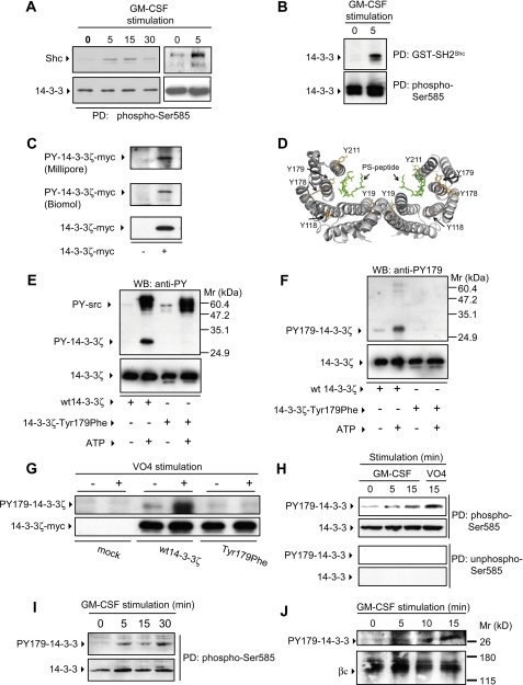
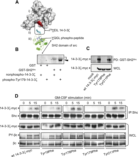
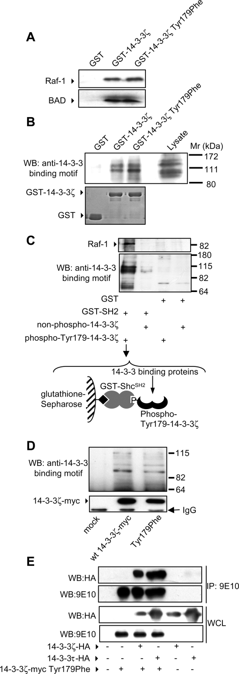
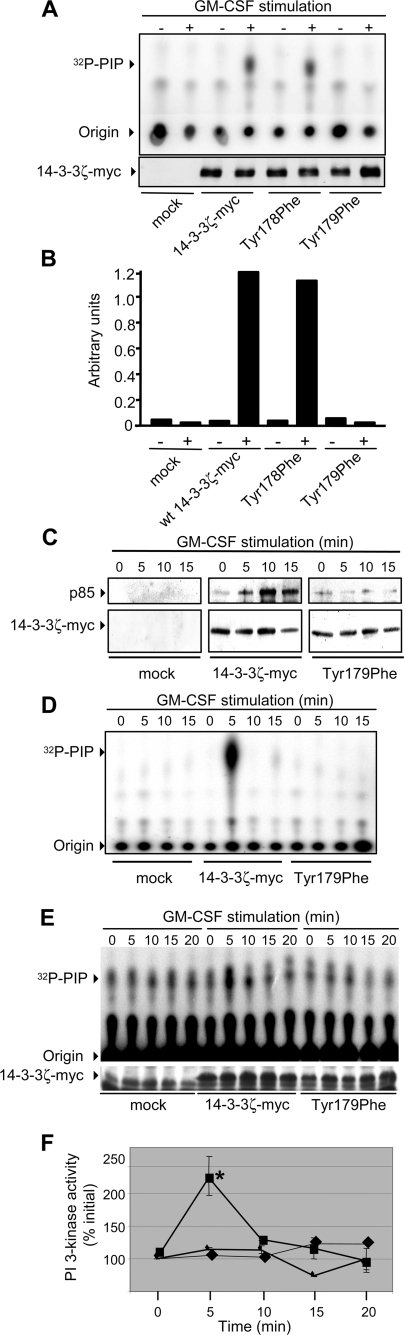
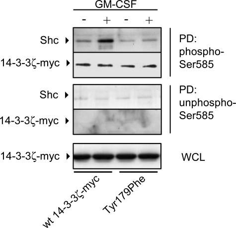
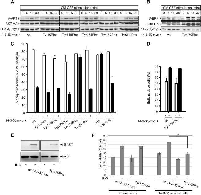
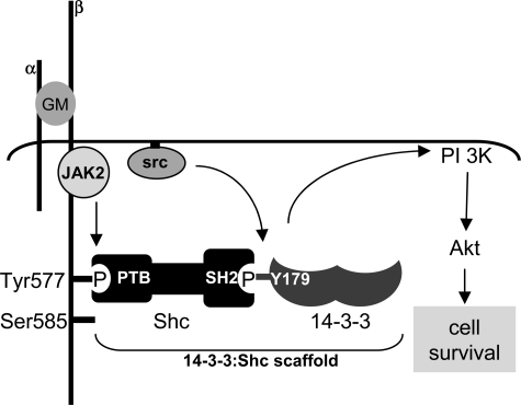
Integrated cascades of protein tyrosine and serine/threonine phosphorylation play essential roles in transducing signals in response to growth factors and cytokines. How adaptor or scaffold proteins assemble signaling complexes through both phosphotyrosine and phosphoserine/threonine residues to regulate specific signaling pathways and biological responses is unclear. We show in multiple cell types that endogenous 14-3-3zeta is phosphorylated on Tyr(179) in response to granulocyte macrophage colony-stimulating factor. Importantly, 14-3-3zeta can function as an intermolecular bridge that couples to phosphoserine residues and also directly binds the SH2 domain of Shc via Tyr(179). The assembly of these 14-3-3:Shc scaffolds is specifically required for the recruitment of a phosphatidylinositol 3-kinase signaling complex and the regulation of CTL-EN cell survival in response to cytokine. The biological significance of these findings was further demonstrated using primary bone marrow-derived mast cells from 14-3-3zeta(-/-) mice. We show that cytokine was able to promote Akt phosphorylation and viability of primary mast cells derived from 14-3-3zeta(-/-) mice when reconstituted with wild type 14-3-3zeta, but the Akt phosphorylation and survival response was reduced in cells reconstituted with the Y179F mutant. Together, these results show that 14-3-3:Shc scaffolds can act as multivalent signaling nodes for the integration of both phosphoserine/threonine and phosphotyrosine pathways to regulate specific cellular responses.


Figures

FIGURE 1.
FIGURE 1.
14-3-3 proteins undergo tyrosine phosphorylation in response to GM-CSF stimulation. A, primary human mononuclear cells from peripheral blood (left panel) or CTL-EN cells (right panel) were stimulated with GM-CSF. Endogenous 14-3-3 was precipitated with a Ser(P)585 peptide (Biotin-NHS-KGGFDFNGPYLGPPHSR(pS)LPDGG; to precipitate total endogenous 14-3-3), and precipitates were immunoblotted with anti-Shc and anti-14-3-3 pAbs. B, factor-dependent CTL-EN cells expressing the human GM-CSF receptor were factor-deprived overnight in medium containing 0.5% FCS and then stimulated with GM-CSF. The cells were lysed, and pulldown experiments were performed using either GST-SH2Shc (to precipitate tyrosine-phosphorylated endogenous 14-3-3) or the Ser(P)585 peptide (to precipitate total endogenous 14-3-3), and precipitates were immunoblotted with anti-14-3-3 pAb. C, HEK 293T cells were transfected with a construct for the expression of wt 14-3-3ζ-Myc. After 48 h, the cells were lysed and 14-3-3ζ-Myc immunoprecipitated using the 9E10 mAb. Immunoprecipitates were subjected to immunoblot analysis with either the 4G10 mAb (Millipore) (top panel), an anti-phosphotyrosine pAb (Biomol) (middle panel) or the 9E10 mAb (bottom panel). D, the crystal structure of human 14-3-3ζ indicates that it is composed of 2 monomers that dimerize to form a large central groove that binds to phosphoserine peptides (PS-peptide) (16). The location of tyrosines 19, 118, 178, 179, and 211 of 14-3-3ζ are indicated. E and F, recombinant purified 14-3-3ζ and 14-3-3ζ-T179F were phosphorylated in vitro using c-Src and then subjected to immunoblot analysis using the 4G10 anti-phosphotyrosine mAb (anti-PY) (E, top panel), anti-Tyr(P)179-14-3-3ζ pAb (anti-PY179) (F, top panel) or anti-14-3-3ζ pAb to indicate loading (E and F, bottom panels). G, HEK-293T cells were transfected with constructs for the expression of wt 14-3-3ζ-Myc or the T179F mutant. After 48 h, the cells were stimulated for 15 min with sodium pervanadate (VO4) (+) or left unstimulated (-). The cells were then lysed and subjected to immunoprecipitation using the 9E10 mAb. Immunoprecipitated proteins were subjected to immunoblot analysis using the anti-Tyr(P)179-14-3-3ζ pAb (G, top panel) and the 9E10 mAb (G, bottom panel). H, CTL-EN cells expressing the human GM-CSF receptor were factor-deprived overnight. The cells were then stimulated with 50 ng/ml GM-CSF or sodium pervanadate (VO4), following which the cells were lysed, and endogenous 14-3-3 was precipitated (PD) using either a Ser(P)585 peptide (Biotin-NHS-KGGFDFNGPYLGPPHSR(pS)LPDGG) or an Ser585 (non-phospho-Ser585) control peptide (Biotin-NHS-KGGFDFNGPYLGPPHSRSLPDGG) adsorbed to streptavidin-Sepharose resin. Pulldowns were subjected to immunoblot analysis using the anti-Tyr(P)179-14-3-3ζ pAb or anti-14-3-3 pAb. I, primary human mononuclear cells were stimulated with GM-CSF and total endogenous 14-3-3 was precipitated as in A. Precipitates were blotted with anti-Tyr(P)179-14-3-3ζ pAb or anti-14-3-3 pAb. J, the UT7 factor-dependent cell line was factor-deprived overnight and then stimulated with 50 ng/ml GM-CSF, following which the cells were lysed, and the βc subunit of the GM-CSF receptor was immunoprecipitated using the 8E4 and 1C1 mAbs. Immunoprecipitates were then subjected to immunoblot analysis with either anti-Tyr(P)179 pAb (top panel) or 1C1 anti-βc mAb (bottom panel). The results are typical of at least two experiments. WB, Western blotting.
FIGURE 2.
FIGURE 2.
Tyr179 of 14-3-3ζ is necessary for GM-CSF-mediated recruitment of Shc. A, structural analysis of 14-3-3ζ and the SH2 domain of Src shows the solvent-accessible surface of 14-3-3ζ (i) and the location of the 179YEIL is indicated as a patch of red on the surface, the 179YEIL motif (cyan) from 14-3-3 in the orientation required to bind to the SH2 domain of Shc (ii), and a cartoon of the SH2 domain of Shc (green) with phosphopeptide (blue) bound (iii). B, purified recombinant GST or GST-SH2Shc bound to glutathione-Sepharose was incubated with either purified recombinant 14-3-3ζ phosphorylated on Tyr179 or a nonphosphorylated 14-3-3ζ control for 1 h in Nonidet P-40 lysis buffer. Pulldowns were washed and subjected to immunoblot analysis with 4G10 mAb. The diffuse gray band corresponds to GST-SH2Shc (bracket), and tyrosine-phosphorylated 14-3-3ζ (arrowhead) lies immediately below. C, HEK 293T cells were transfected with constructs expressing either the wt 14-3-3ζ-Myc or the indicated mutants. After 48 h, the cells were lysed, and lysates were incubated with either the GST-SH2Shc or a GST control. Pulldowns (PD) and whole cell lysates (WCL) were subjected to immunoblot analysis using the 9E10 mAb. D, CTL-EN cells expressing the human GM-CSF receptor were electroporated with constructs for the expression of either wt 14-3-3ζ-Myc or the indicated mutants and factor-deprived overnight. The cells were then stimulated with 50 ng/ml GM-CSF followed by lysis and anti-Shc immunoprecipitation. Immunoprecipitates (IP) were subjected to immunoblot analysis using the 9E10 mAb or anti-Shc pAb. Tyrosine phosphorylation of the βc subunit of the GM-CSF receptor in response to GM-CSF was examined in lysates (WCL) using the 4G10 mAb (PYc). WCL were also immunoblotted using the 1C1 anti-βc mAb and the 9E10 mAb to demonstrate equal loading. The results are typical of two experiments.
FIGURE 3.
FIGURE 3.
The 14-3-3ζ-T179F mutant is able to bind phosphoserine/threonine target proteins and heterodimerize with other isoforms of 14-3-3. HEK 293T cells were lysed and whole cell lysates subjected to pulldown experiments using either GST-Sepharose, GST-14-3-3ζ-Sepharose, or GST-14-3-3ζ T179F-Sepharose. Pulldowns were examined by immunoblot analysis using anti-Raf-1 and anti-BAD pAb (A) or a phospho-specific antibody that recognizes phosphorylated 14-3-3 binding motifs (Cell Signaling) (B). C, recombinant purified 14-3-3ζ was phosphorylated with c-Src (Tyr(P)179-14-3-3ζ) or left nonphosphorylated (non-phospho-14-3-3ζ) and then incubated with either GST-SH2Shc or GST alone bound to glutathione-Sepharose resin. The resin was washed to remove unbound 14-3-3ζ, and the assembled complexes (the complex assembled in lane 1 and used in the pulldown experiment is illustrated) were then used to pulldown 14-3-3-binding proteins from HEK 293T cell lysates. The pulldowns were washed in Nonidet P-40 lysis buffer and subjected to immunoblot analysis with phospho-specific antibodies that recognize phosphorylated 14-3-3 binding motifs or anti-Raf-1 pAb. D, HEK-293T cells were either mock transfected or transfected with constructs for the expression of wt 14-3-3ζ-Myc or the T179F mutant. After 48 h, the cells were lysed in Nonidet P-40 lysis buffer and subjected to immunoprecipitation using the 9E10 mAb. Immunoprecipitated proteins were subjected to SDS-PAGE and immunoblot analysis with phospho-specific antibodies that recognize phosphorylated 14-3-3 binding motifs (top panel) or the 9E10 mAb (bottom panel). E, HEK-293T cells were co-transfected with the indicated combinations of 14-3-3 isoforms. After 48 h, the cells were lysed, and lysates were immunoprecipitated (IP) with the 9E10 mAb. Immunoprecipitates and whole cell lysates (WCL) were subjected to SDS-PAGE and immunoblotted using the 9E10 mAb or the 12CA5 mAb. The 14-3-3ζ-Myc-T179F mutant was able to co-immunoprecipitate with 14-3-3γ-EE (data not shown), 14-3-3ζ-HA, and 14-3-3τ-HA. WB, Western blotting.
FIGURE 4.
FIGURE 4.
Tyr179 of 14-3-3ζ is necessary for the GM-CSF-mediated recruitment and activation of PI 3-kinase. A, CTL-EN cells expressing the human GM-CSF receptor were electroporated with constructs for the expression of either the wt 14-3-3ζ-Myc or the indicated mutants, and cells were factor-deprived overnight. The cells were then stimulated with 50 ng/ml GM-CSF for 5 min and lysed, and 14-3-3ζ-Myc was immunoprecipitated using the 9E10 mAb. Immunoprecipitates were then washed, and PI 3-kinase activity was measured in vitro using 32P-γATP and phosphatidylinositol (PIP) as substrates. Extracted 32P-lipids were subjected to thin layer chromatography, and indicated are 32P-PIP and the origin. Quantification of these PhosphorImager signals (Typhoon) is shown in B. C, CTL-EN cells were electroporated without plasmid (mock), wt 14-3-3ζ-Myc, or the T179F mutant, following which the cells were factor-deprived and stimulated with GM-CSF as for A. 14-3-3ζ-Myc was immunoprecipitated with the 9E10 mAb with ⅓ of the immunoprecipitate used for PI 3-kinase assays (D) and 2/3 used for immunoblot analysis with anti-p85 pAb or 9E10 mAb (C). E, UT7 factor-dependent cells were electroporated with the indicated plasmids and PI 3-kinase assays were performed as in A and D. Quantification of PhosphorImager signals from two experiments is shown, and the error bars indicate the standard deviations (F). PI 3-kinase activity associated with the T179F mutant (♦) at 5 min was significantly decreased in comparison to the activity associated with wt 14-3-3ζ (▪) (*, p < 0.03). Mock transfected cells are indicated by the ▴.
FIGURE 5.
FIGURE 5.
14-3-3ζ can simultaneously bind phosphoserine residues and Shc. CTL-EN cells were electroporated with either wt 14-3-3ζ-Myc or the T179F mutant following which the cells were factor-deprived overnight and then stimulated with 50 ng/ml GM-CSF for 5 min. The cells were then lysed and pulldowns (PD) were performed using either a Ser(P)585 peptide (Biotin-NHS-KGGFDFNGPYLGPPHSR(pS)LPDGG) or a non-phospho-Ser585 control peptide (Biotin-NHS-KGGFDFNGPYLGPPHSRSLPDGG) adsorbed to streptavidin-Sepharose. PD and whole cell lysates (WCL) were subjected to SDS-PAGE and immunoblot analysis using anti-Shc pAb and the 9E10 mAb. The results are typical of two experiments.
FIGURE 6.
FIGURE 6.
Tyr179 of 14-3-3ζ required for the phosphorylation of Akt and is necessary and sufficient for regulating GM-CSF-mediated cell survival. CTL-EN cells expressing the GM-CSF receptor were co-electroporated with constructs expressing Akt-HA (A) or ERK-HA (B) and either wt 14-3-3ζ-Myc or the indicated 14-3-3ζ mutants. Co-transfection of Akt-HA or ERK-HA reporter plasmids allowed the specific examination of Akt and ERK phosphorylation in response to GM-CSF in transfected cells only. The cells were factor-deprived overnight and then stimulated with GM-CSF. The cells were then lysed, and the lysates were subjected to immunoblot analysis using the anti-phospho-Akt pAb (Thr308 and Ser473) (A), anti-active-ERK pAb (B) or the 9E10 or 12CA5 mAb. C, CTL-EN cells expressing the βcS585G mutant of GM-CSF receptor were co-electroporated with constructs expressing EGFP (to allow detection of transfected cells) and either wt 14-3-3ζ-Myc or the indicated mutants. After 24 h, the cells were washed and plated in medium containing either no factor (-) or 50 ng/ml GM-CSF (+). After a further 48 h, the cells were stained with annexin V-PE and analyzed by flow cytometry. Shown is the percentage of EGFP-positive cells that are annexin V-PE-positive. Cell proliferation was also examined (D). CTL-EN cells expressing the βcS585G mutant of GM-CSF receptor were co-electroporated with constructs expressing enhanced cyan fluorescent protein and either wt 14-3-3ζ-Myc or the T179F mutant. After 24 h, cyan fluorescent protein-positive cells were purified by fluorescence-activated cell sorting and plated in either no factor (-) or 50 ng/ml GM-CSF (+) for 24 h with a BrdUrd pulse for the last 4 h. The cells were then fixed, stained for BrdUrd incorporation, and analyzed by flow cytometry. E, primary bone marrow-derived mast cells from 14-3-3ζ-/- mice were electroporated with either wt 14-3-3ζ-Myc or the T179F mutant. After 24 h, the cells were washed and plated at 1.5 × 10⁵/ml in serum-free DMEM containing 0.1% bovine serum albumin overnight and then stimulated with 1000 pm IL-3 and Akt phosphorylation was examined as in A. F, primary bone marrow-derived mast cells from wt and 14-3-3ζ-/- mice were electroporated with either wt 14-3-3ζ-Myc or the T179F mutant together with EGFP to allow identification of transfected cells. After 24 h, the cells were washed and plated at 1.5 × 10⁵/ml in serum-free DMEM containing 0.1% bovine serum albumin and 1000 pm IL-3. The number of viable GFP-positive cells was determined by flow cytometry and forward/side scatter characteristics of mast cells at 48 h as a percentage of the initial GFP-positive population to account for differences in transfection efficiency. A significant reduction in IL-3-mediated survival of 14-3-3ζ-/- bone marrow-derived mast cells expressing the T179F mutant was observed when compared with knock-out cells expressing the wt 14-3-3ζ-Myc (*, p < 0.0007 from two separate experiments). All of the results are representative of at least two experiments.
FIGURE 7.
FIGURE 7.
Model for the assembly of 14-3-3:Shc scaffolds. Stimulation with 50 ng/ml GM-CSF (GM) results in the activation of tyrosine kinases including JAK2 and Src family of kinases. Although we have shown that the activated receptor constitutes a high order dodecamer (33), only two subunits of the receptor are illustrated. JAK2 can phosphorylate Tyr577 of βc, whereas Src family kinases can phosphorylate Tyr179 of 14-3-3ζ. The phosphorylation of 14-3-3ζ on Tyr179 provides a docking site for the direct binding of the SH2 domain of Shc. The 14-3-3:Shc scaffold bound to Tyr577 of the βc subunit of the GM-CSF receptor is specifically required for the activation of the PI 3-kinase/Akt pathway and the regulation of cell survival.

References

    1. Pawson, T., and Scott, J. D. (1997) Science 278 2075-2080 - PubMed
    1. Pawson, T. (2004) Cell 116 191-203 - PubMed
    1. Songyang, Z., Shoelson, S. E., McGlade, J., Olivier, P., Pawson, T., Bustelo, X. R., Barbacid, M., Sabe, H., Hanafusa, H., and Yi, T. (1994) Mol. Cell Biol. 14 2777-2785 - PMC - PubMed
    1. Yaffe, M. B. (2002) FEBS Lett. 513 53-57 - PubMed
    1. Aitken, A. (2006) Semin. Cancer Biol. 16 162-172 - PubMed

Publication types

MeSH terms

Substances