. 2009 Mar;174(3):1027-36.
doi: 10.2353/ajpath.2009.080501. Epub 2009 Feb 13.

Spontaneous metastasis of prostate cancer is promoted by excess hyaluronan synthesis and processing

Affiliations

Spontaneous metastasis of prostate cancer is promoted by excess hyaluronan synthesis and processing

Alamelu G Bharadwaj et al. Am J Pathol. 2009 Mar.

Abstract

Accumulation of extracellular hyaluronan (HA) and its processing enzyme, the hyaluronidase Hyal1, predicts invasive, metastatic progression of human prostate cancer. To dissect the roles of hyaluronan synthases (HAS) and Hyal1 in tumorigenesis and metastasis, we selected nonmetastatic 22Rv1 prostate tumor cells that overexpress HAS2, HAS3, or Hyal1 individually, and compared these cells with co-transfectants expressing Hyal1 + HAS2 or Hyal1 + HAS3. Cells expressing only HAS were less tumorigenic than vector control transfectants on orthotopic injection into mice. In contrast, cells co-expressing Hyal1 + HAS2 or Hyal1 + HAS3 showed greater than sixfold and twofold increases in tumorigenesis, respectively. Fluorescence and histological quantification revealed spontaneous lymph node metastasis in all Hyal1 transfectant-implanted mice, and node burden increased an additional twofold when Hyal1 and HAS were co-expressed. Cells only expressing HAS were not metastatic. Thus, excess HA synthesis and processing in concert accelerate the acquisition of a metastatic phenotype by prostate tumor cells. Intratumoral vascularity did not correlate with either tumor size or metastatic potential. Analysis of cell cycle progression revealed shortened doubling times of Hyal1-expressing cells. Both adhesion and motility on extracellular matrix were diminished in HA-overproducing cells; however, motility was increased twofold by Hyal1 expression and fourfold to sixfold by Hyal1/HAS co-expression, in close agreement with observed metastatic potential. This is the first comprehensive examination of these enzymes in a relevant prostate cancer microenvironment.


Figures
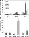
Figure 1
Figure 1
Orthotopic prostate tumorigenesis is enhanced by co-expression of HA-synthesizing and HA-turnover enzymes. Single cell suspensions of transfected cell lines as indicated were injected in 10 μl of serum-free RPMI to the dorsal prostate of male NOD/SCID mice (seven per group, repeated in triplicate). A: Tumor growth was monitored longitudinally at 2-week intervals by near infrared optical imaging. Mice were imaged 72 hours after an intravenous injection of IRDye 800CW EGF (1 nmol). Images were analyzed for total tumor fluorescence detected at signal to noise ratios of >5 SDs above background. B: At 6 weeks after surgery, after the final image capture, animals were sacrificed and tumors were harvested and weighed. Plotted is the mean tumor wet weight ± SEM for all mice; *P < 0.01.
Figure 2
Figure 2
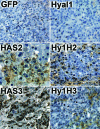
HA content of orthotopic tumor sections. Formalin-fixed, paraffin-embedded tumors were sectioned and stained with biotinylated HA binding protein, followed by detection with streptavidin-conjugated HRP and diaminobenzidine precipitation to visualize HA content. Sections were counterstained with hematoxylin and representative fields were digitally photographed at ×200 magnification.
Figure 3
Figure 3
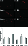
Enhanced tumorigenesis in the prostate does not correlate with intratumoral blood vessel density. Orthotopic tumors were embedded in OCT compound and cryosectioned. Sections were stained with anti-CD31-PE conjugate and 10 randomly selected sections per tumor were digitally photographed under a fluorescence microscope at ×400 magnification with a 5-second exposure time. Representative images from each tumor type as indicated are shown. Images were processed as indicated in Materials and Methods to obtain total vessel area per section as pixel density. Mean pixel density per section ± SEM is plotted for 10 sections of three different tumors per group; *P < 0.01, **P < 0.05.
Figure 4
Figure 4
Spontaneous lymph node metastasis is initiated by Hyal1 and potentiated by HAS/Hyal1 co-expression. Three days before study termination, mice were injected intravenously with IRDye 800CW EGF (1 nmol). After final image capture, animals were sacrificed and dissected to expose the para-aortic lymph nodes. A: Images of lymph nodes in situ from representative animals are shown for comparison at the same look-up table. LN indicates position of lymph nodes; arrows indicate fluorescence in the lymph nodes. Excised lymph nodes from all animals in the study were imaged intact for fluorescence quantification (B, D) and sectioned for histology (C, E). Nodes shown are from animals injected with HAS2 (B, C) and Hyal1/HAS2 (D, E) transfectants. F: Fluorescence intensity (800 channel signal with signal-to-noise ratio >3) of all excised nodes was normalized to fluorescence of nodes from mice injected with control GFP transfectants. Mean ± SEM is plotted; *P < 0.01.
Figure 5
Figure 5
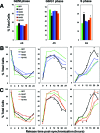
Cell-cycle analysis reveals accelerated cell cycling of Hyal1-overexpressing cells. Transfectant cell lines were fixed, permeabilized, propidium iodide-stained, and analyzed by flow cytometry. A: Respective mean percentages of cells in each phase of the cycle are plotted ± SEM for a total of three experiments in asynchronous cultures. Cells were synchronized in the G2/M phase by nocodazole treatment, then released by replacement with standard medium for the indicated times and similarly analyzed by flow cytometry. Mean ± SEM of triplicate assays is plotted for each phase of the cell cycle at each time point as indicated at the top of each column; *P < 0.01. Plots compare HAS2 (dark blue triangles), Hyal1 (black squares), and Hyal1/HAS2 (light blue circles) cells (B) or HAS3 (red triangles), Hyal1 (black squares), and Hyal1/HAS3 (orange circles) cells (C).
Figure 6
Figure 6
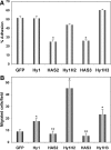
Cell adhesion and migration to collagen are impaired by HAS expression but migration is increased synergistically with Hyal1 and HAS co-expression. A: Cell adhesion to type IV collagen (5 μg/ml) was quantified in a precoated 96-well plate, to which calcein-labeled tumor cell suspensions were added. Nonadherent cells were removed by gentle washing after a 2-hour incubation at 37°C. Remaining adherent cells were lysed and the fluorescence of each well was normalized to a standard curve for each cell line. Results plotted are the mean ± SEM of quadruplicate wells from a total of three experiments; *P < 0.01. B: Cell migration was assayed in a modified Boyden chamber. Stably transfected 22Rv1 cells as indicated were trypsin-released and resuspended in serum-free RPMI. The lower wells of the chamber contained type IV collagen (25 μmol/L) in serum-free RPMI. Cells (20,000/well) were placed in quadruplicate wells of the upper chamber, separated from the lower wells by a polycarbonate membrane with 8 μm pore size. After 20 hours of incubation at 37°C, membranes were fixed and stained. Migrated cells were counted in five random fields per well at ×150 magnification. Average number of cells per field is plotted ± SEM for each cell line; *P < 0.01, **P < 0.05.

References

    1. Pienta KJ, Smith DC. Advances in prostate cancer chemotherapy: a new era begins. CA Cancer J Clin. 2005;55:300–318. - PubMed
    1. Fraser JR, Laurent TC, Laurent UB. Hyaluronan: its nature, distribution, functions and turnover. J Intern Med. 1997;242:27–33. - PubMed
    1. Stern R. Hyaluronan metabolism: a major paradox in cancer biology. Pathol Biol (Paris) 2005;53:372–382. - PubMed
    1. Toole BP. Hyaluronan promotes the malignant phenotype. Glycobiology. 2002;12:37R–42R. - PubMed
    1. Toole BP. Hyaluronan: from extracellular glue to pericellular cue. Nat Rev Cancer. 2004;4:528–539. - PubMed

Publication types

MeSH terms

Substances