. 2009 Apr;191(8):2711-20.
doi: 10.1128/JB.01832-08. Epub 2009 Feb 13.

SleC is essential for cortex peptidoglycan hydrolysis during germination of spores of the pathogenic bacterium Clostridium perfringens

Affiliations

SleC is essential for cortex peptidoglycan hydrolysis during germination of spores of the pathogenic bacterium Clostridium perfringens

Daniel Paredes-Sabja et al. J Bacteriol. 2009 Apr.

Abstract

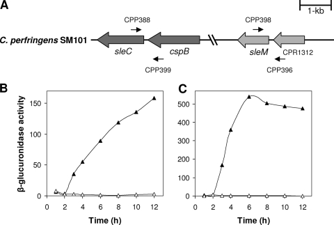
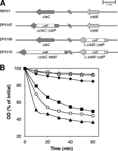
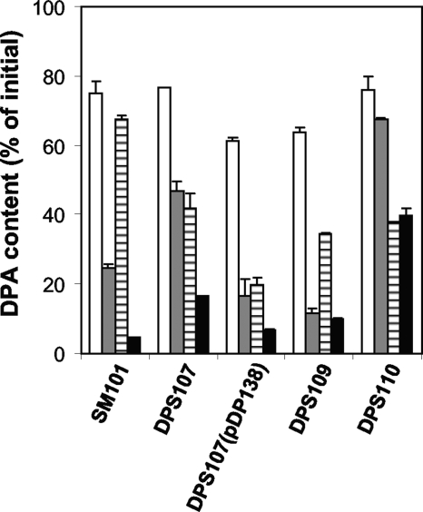
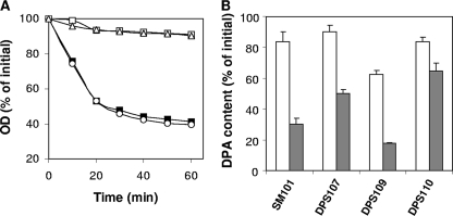
Clostridial spore germination requires degradation of the spore's peptidoglycan (PG) cortex by cortex-lytic enzymes (CLEs), and two Clostridium perfringens CLEs, SleC and SleM, degrade cortex PG in vitro. We now find that only SleC is essential for cortex hydrolysis and viability of C. perfringens spores. C. perfringens sleC spores did not germinate completely with nutrients, KCl, or a 1:1 chelate of Ca(2+) and dipicolinic acid (Ca-DPA), and the colony-forming efficiency of sleC spores was 10(3)-fold lower than that of wild-type spores. However, sleC spores incubated with various germinants released most of their DPA, although slower than wild-type or sleM spores, and DPA release from sleC sleM spores was very slow. In contrast, germination and viability of sleM spores were similar to that of wild-type spores, although sleC sleM spores had 10(5)-fold-lower viability. These results allow the following conclusions about C. perfringens spore germination: (i) SleC is essential for cortex hydrolysis; (ii) although SleM can degrade cortex PG in vitro, this enzyme is not essential; (iii) action of SleC alone or with SleM can accelerate DPA release; and (iv) Ca-DPA does not trigger spore germination by activation of CLEs.


Figures

FIG. 1.
FIG. 1.
(A to C) Arrangement and expression of sle genes in C. perfringens SM101. (A) The arrangement of sleC and sleM in C. perfringens SM101 is shown, and the locations of the primers used to amplify the upstream regions of each gene are indicated. The sleC and sleM promoters were predicted to be within the intergenic regions between sleC and cspB and between sleM and CPR1312, respectively (18). (B and C) GUS specific activities from sleC-gusA (B) and sleM-gusA (C) fusions in C. perfringens SM101 grown in TGY vegetative (▵) and DS sporulation (▴) media were determined as described in Materials and Methods. The data represent averages of three independent experiments, and time zero denotes the time of inoculation of cells into either TGY or DS medium.
FIG. 2.
FIG. 2.
(A and B) C. perfringens sle deletion mutants and germination of their spores in BHI broth. (A) Arrangement of sleC and sleM in various C. perfringens deletion mutant strains. (B) Spores of C. perfringens strains SM101 (wild-type) (▪), DPS107 (sleC) (▵), DPS109 (sleM) (○), DPS110 (sleC sleM) (□), and DPS107(pDP138) (sleC strain complemented with wild-type sleC) (▴) were incubated at 40°C in BHI broth, and the OD600 was measured as described in Materials and Methods. Spores of C. perfringens strains SM101 (wild-type) (⧫) and DPS107 (sleC) were incubated at 40°C in 25 mM sodium phosphate buffer (pH 7.0), and the OD600 was measured as described in Materials and Methods. There was essentially no decrease in the OD600 for the sleC spores incubated in buffer alone (data not shown).
FIG. 3.
FIG. 3.
(A and B) Germination of C. perfringens spores over long periods on BHI agar plates. (A) Spores of C. perfringens strains DPS107 (sleC) (□) and DPS110 (sleC sleM) (░⃞) were applied to BHI plates that were incubated at 37°C for 7 days, and the total number of colonies were counted every 24 h and expressed as CFU. (B) The plates described in panel A were photographed every 24 h. Similar results in the experiments in panels A and B were obtained with two different batches of spores, and error bars indicate the standard deviations.
FIG. 4.
FIG. 4.
DPA release during the germination of C. perfringens spores with BHI broth. Heat-activated spores of strains SM101 (wild-type), DPS107 (sleC), DPS107(pDP138) (sleC strain complemented with wild-type sleC), DPS109 (sleM), and DPS110 (sleC sleM) were incubated at 40°C in BHI broth and, after 1 h (░⃞) and 18 h (▪), DPA release was measured as described in Materials and Methods. C. perfringens spores from various strains were also incubated in 25 mM sodium phosphate buffer (pH 7.0), and after 1 (□) or 18 h (▤) the DPA release was measured as described in Materials and Methods. The data represent the average of two independent experiments with two different spore preparations, and error bars indicate the standard deviations.
FIG. 5.
FIG. 5.
(A to C) Germination of spores of C. perfringens strains with KCl. (A) Heat-activated C. perfringens spores of strains SM101(wild-type) (▪), DPS107 (sleC) (▵), DPS109 (sleM) (○), and DPS110 (sleC sleM) (□) were germinated with KCl, and the OD600 was measured as described in Materials and Methods. Heat-activated spores of strain SM101 (wild-type) (⧫) were also incubated in 25 mM sodium phosphate buffer (pH 7.0) alone, and the OD600 was measured. (B) DPA release during C. perfringens spore germination with KCl. Heat-activated spores of C. perfringens strains were germinated with KCl, and after 1 h (□) and 18 h (░⃞) the DPA content of the spores was measured as described in Materials and Methods. DPA release of C. perfringens spores from various strains in 25 mM sodium phosphate buffer (pH 7.0) was as shown in Fig. 4. (C) Release of hexosamine-containing material during C. perfringens spore germination with KCl. Heat-activated spores of C. perfringens strains were germinated with KCl and, after 2 h, the hexosamine-containing material released into the medium was measured as described in Materials and Methods. Values for hexosamine-containing material released are expressed relative to the amount of hexosamine in dormant spores that was defined as 100%. The data represent the average of two independent experiments with two different spore preparations, and error bars indicate the standard deviations.
FIG. 6.
FIG. 6.
(A and B) Germination of C. perfringens spores with Ca-DPA. (A) Heat-activated C. perfringens spores of strains SM101 (wild-type) (▪), DPS107 (sleC) (▵), DPS109 (sleM) (○), and DPS110 (sleC sleM) (□) were germinated with 50 mM Ca-DPA, and the OD600 was measured as described in Materials and Methods. (B) Heat-activated spores of C. perfringens strains were germinated with 50 mM Ca-DPA (░⃞) and 25 mM Tris-HCl buffer (pH 8.0) (□) for 1 h, and the DPA remaining in the spores was measured as described in Materials and Methods. The data represent the average of two independent experiments with two different spore preparations, and error bars indicate the standard deviations.
FIG. 7.
FIG. 7.
Dodecylamine germination of C. perfringens spores. C. perfringens spores of strains SM101 (wild-type) (▪), DPS107 (sleC) (▵), DPS109 (sleM) (○), DPS110 (sleC sleM) (□), and DPS107(pDP138) (sleC strain complemented with wild-type sleC) (▴) were germinated with dodecylamine, and at various times the DPA release was measured as described in Materials and Methods.

References

    1. Atluri, S., K. Ragkousi, D. E. Cortezzo, and P. Setlow. 2006. Cooperativity between different nutrient receptors in germination of spores of Bacillus subtilis and reduction of this cooperativity by alterations in the GerB receptor. J. Bacteriol. 18828-36. - PMC - PubMed
    1. Atrih, A., P. Zollner, G. Allmaier, and S. J. Foster. 1996. Structural analysis of Bacillus subtilis 168 endospore peptidoglycan and its role during differentiation. J. Bacteriol. 1786173-6183. - PMC - PubMed
    1. Bagyan, I., and P. Setlow. 2002. Localization of the cortex lytic enzyme CwlJ in spores of Bacillus subtilis. J. Bacteriol. 1841219-1224. - PMC - PubMed
    1. Boland, F. M., A. Atrih, H. Chirakkal, S. J. Foster, and A. Moir. 2000. Complete spore-cortex hydrolysis during germination of Bacillus subtilis 168 requires SleB and YpeB. Microbiology 14657-64. - PubMed
    1. Bruggemann, H., S. Baumer, W. F. Fricke, A. Wiezer, H. Liesegang, I. Decker, C. Herzberg, R. Martinez-Arias, R. Merkl, A. Henne, and G. Gottschalk. 2003. The genome sequence of Clostridium tetani, the causative agent of tetanus disease. Proc. Natl. Acad. Sci. USA 1001316-1321. - PMC - PubMed

Publication types

MeSH terms

LinkOut - more resources