. 2009 Jun;191(11):3451-61.
doi: 10.1128/JB.01539-08. Epub 2009 Feb 13.

The Escherichia coli common pilus and the bundle-forming pilus act in concert during the formation of localized adherence by enteropathogenic E. coli

Affiliations

The Escherichia coli common pilus and the bundle-forming pilus act in concert during the formation of localized adherence by enteropathogenic E. coli

Zeus Saldaña et al. J Bacteriol. 2009 Jun.

Abstract

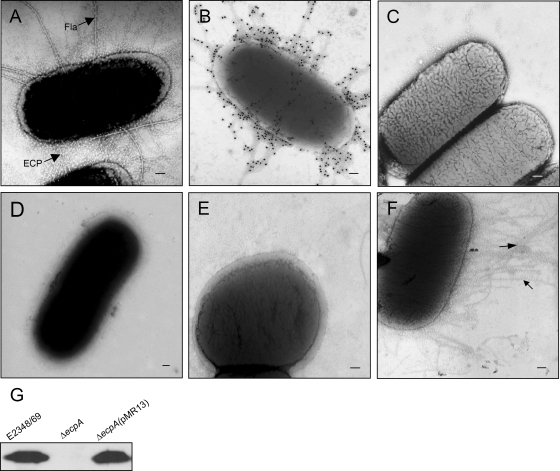
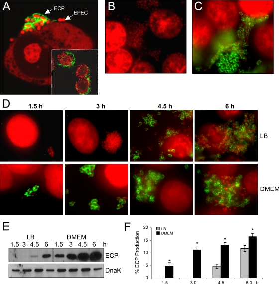
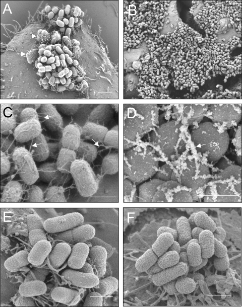
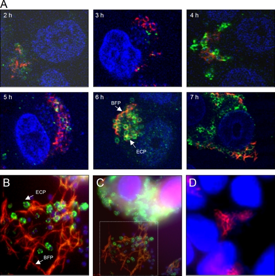
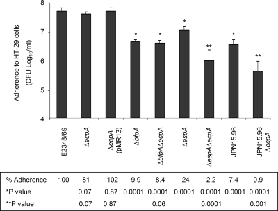
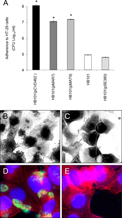
Although the bundle-forming pilus (BFP) of enteropathogenic Escherichia coli (EPEC) mediates microcolony formation on epithelial cells, the adherence of BFP-deficient mutants is significantly abrogated, but the mutants are still adherent due to the presence of intimin and possibly other adhesins. In this study we investigated the contribution of the recently described E. coli common pilus (ECP) to the overall adherence properties of EPEC. We found that ECP and BFP structures can be simultaneously observed in the course (between zero time and 7 h during infection) of formation of localized adherence on cultured epithelial cells. These two pilus types colocalized at different levels of the microcolony topology, tethering the adhering bacteria. No evidence of BFP disappearance was found after prolonged infection. When expressed from a plasmid present in nonadherent E. coli HB101, ECP rendered this organism highly adherent at levels comparable to those of HB101 expressing the BFP. Purified ECP bound in a dose-dependent manner to epithelial cells, and the binding was blocked with anti-ECP antibodies, confirming that the pili possess adhesin properties. An ECP mutant showed only a modest reduction in adherence to cultured cells due to background expression levels of BFP and intimin. However, isogenic mutants not expressing EspA or BFP were significantly less adherent when the ecpA gene was also deleted. Furthermore, a DeltaespA DeltaecpA double mutant (unable to translocate Tir and to establish intimate adhesion) was at least 10-fold less adherent than the DeltaespA and DeltaecpA single mutants, even in the presence of BFP. A Delta bfp DeltaespA DeltaecpA triple mutant showed the least adherence compared to the wild type and all the isogenic mutant strains tested, suggesting that ECP plays a synergistic role in adherence. Our data indicate that ECP is an accessory factor that, in association with BFP and other adhesins, contributes to the multifactorial complex interaction of EPEC with host epithelial cells.


Figures

FIG. 1.
FIG. 1.
ECP on EPEC as shown by electron microscopy and immunogold labeling. (A) Electron micrograph of E2348/69 grown in DMEM displaying abundant peritrichous fine pili (ECP) and flagella (Fla). (B and C) Immunogold labeling of ECP on E2348/69 with anti-ECP antibodies and preimmune serum, respectively. (D) E2348/69 grown in LB medium showing no pili. (E) E2348/69ΔecpA mutant showing no ECP. (F) E2348/69ΔecpA(pMR13) with restored ECP production. Scale bars, 100 nm. (G) Detection of EcpA in normalized HCl-treated whole-cell extracts.
FIG. 2.
FIG. 2.
ECP on EPEC strains adhering to cultured epithelial cells and kinetics of ECP production. (A) E2348/69 adhering to cultured epithelial cells after 6 h of incubation and reaction with anti-ECP antibodies and Alexa Fluor 488-conjugated secondary antibody (green). Cellular and bacterial DNA was stained with propidium iodide (red). The inset shows a lower magnification of an EPEC microcolony (red) with bacteria producing ECP (green). (B) E2348/69ΔecpA mutant. (C) E2348/69ΔecpA(pMR13). (D) Kinetics of ECP production by bacteria pregrown overnight in LB medium or DMEM. (E) Detection of EcpA in normalized HCl-treated whole-cell extracts. Detection of DnaK with anti-DnaK antibody was used as a loading control. (F) Level of ECP as determined by flow cytometry in bacteria adhering to epithelial cells. The data are representative data from two experiments performed in triplicate. The asterisk indicates that there was a statistically significant difference compared with LB medium-grown bacteria.
FIG. 3.
FIG. 3.
High-resolution SEM analysis of EPEC E2348/69 adhering to HT-29 cells. (A) At 3 h postinfection EPEC forms a typical LA microcolony and AE lesions manifested by pedestals with bacteria on top (arrows). Scale bar, 2 μm. (B) At 6 h postinfection the microcolony has spread throughout the cell monolayer. Scale bar, 6 μm. (C) High magnification of panel B, showing abundant peritrichous thin fibers (arrows) that tether the bacteria together. Scale bar, 1 μm. (D) Immuno-SEM gold labeling of ECP protruding from bacteria with anti-ECP antibodies and secondary rabbit IgG conjugated to 30-nm gold particles. Scale bar, 1 μm (E) SEM analysis of E2348/69ΔecpA mutant. (F) Immuno-SEM gold labeling of E2348/69ΔecpA with anti-ECP antibodies. Scale bar, 1 μm.
FIG. 4.
FIG. 4.
Simultaneous production of ECP and BFP during microcolony formation by E2348/69. (A) Kinetics (2 to 7 h) of ECP (green) and BFP (red) expression in the presence of HT-29 cells using confocal microscopy. Cellular and bacterial DNA was stained blue with DAPI. ECP are tightly associated with the bacterial surface, while the BFP extend out from the bacteria throughout the cluster. The confocal microscopy micrographs were taken at a magnification of ×60. (C) IFM detection of ECP and BFP after 6 h of infection. (B) Magnification of the framed area in panel C. Long and thick BFP structures are evident at this time point. (D) E2348/69ΔecpA mutant.
FIG. 5.
FIG. 5.
Contribution of ECP to EPEC adherence compared with the contributions of other adhesins. The levels of adherence to HT-29 cells of wild-type and mutant strains were determined by plating serial dilutions of adhering bacteria. The adherence experiments were repeated at least three times in triplicate on separate days, and the bars indicate the means of the averages of the results obtained in the three experiments performed. The error bars indicate the standard deviations for the averages of all the results obtained in the three experiments performed. The levels of adherence and the P values are indicated below the graph. A statistical analysis was done using a paired Student t test. *, statistically significant compared with the wild-type strain; **, statistically significant compared with the wild-type strain and the parental strain.
FIG. 6.
FIG. 6.
Production of ECP confers a high level of adherence to nonadherent E. coli HB101. (A) Bacteria were pregrown in DMEM containing 0.1 mM IPTG to induce the expression of the ecpAB genes in HB101(pMAT9), and the adherence assay was performed for 6 h. *, P < 0.0001. (B and C) Light microscopy micrographs comparing the adherence of HB101(pMAT9) and of HB101, respectively, to HeLa cells (magnification, ×60). (D) Immunofluorescence showing high levels of ECP expression by HB101 carrying pMAT9. (E) Negative control strain HB101 showing no fluorescence (magnification, ×60). The adherence experiments were repeated at least three times in triplicate on separate days, and the bars indicate the means of the averages of the results obtained in the three experiments performed. The error bars indicate the standard deviations for the averages of all the results obtained in the three experiments performed.

References

    1. Baldini, M. M., J. B. Kaper, M. M. Levine, D. C. Candy, and H. W. Moon. 1983. Plasmid-mediated adhesion in enteropathogenic Escherichia coli. J. Pediatr. Gastroenterol. Nutr. 2534-538. - PubMed
    1. Bieber, D., S. W. Ramer, C. Y. Wu, W. J. Murray, T. Tobe, R. Fernandez, and G. K. Schoolnik. 1998. Type IV pili, transient bacterial aggregates, and virulence of enteropathogenic Escherichia coli. Science 2802114-2118. - PubMed
    1. Blattner, F. R., G. Plunkett III, C. A. Bloch, N. T. Perna, V. Burland, M. Riley, J. Collado-Vides, J. D. Glasner, C. K. Rode, G. F. Mayhew, J. Gregor, N. W. Davis, H. A. Kirkpatrick, M. A. Goeden, D. J. Rose, B. Mau, and Y. Shao. 1997. The complete genome sequence of Escherichia coli K-12. Science 2771453-1474. - PubMed
    1. Brinton, C. C., Jr. 1959. Non-flagellar appendages of bacteria. Nature 183782-786. - PubMed
    1. Cleary, J., L. C. Lai, R. K. Shaw, A. Straatman-Iwanowska, M. S. Donnenberg, G. Frankel, and S. Knutton. 2004. Enteropathogenic Escherichia coli (EPEC) adhesion to intestinal epithelial cells: role of bundle-forming pili (BFP), EspA filaments and intimin. Microbiology 150527-538. - PubMed

Publication types

MeSH terms