. 2009 Feb;21(2):429-41.
doi: 10.1105/tpc.108.064477. Epub 2009 Feb 13.

Nuclear activity of ROXY1, a glutaredoxin interacting with TGA factors, is required for petal development in Arabidopsis thaliana

Affiliations

Nuclear activity of ROXY1, a glutaredoxin interacting with TGA factors, is required for petal development in Arabidopsis thaliana

Shutian Li et al. Plant Cell. 2009 Feb.

Erratum in

  • Plant Cell. 2014 Mar;26(3):1378
  • CORRECTION.
    [No authors listed] [No authors listed] Plant Cell. 2018 Jan;30(1):249-252. doi: 10.1105/tpc.17.00893. Epub 2018 Jan 12. Plant Cell. 2018. PMID: 29330201 Free PMC article. No abstract available.

Abstract

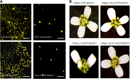
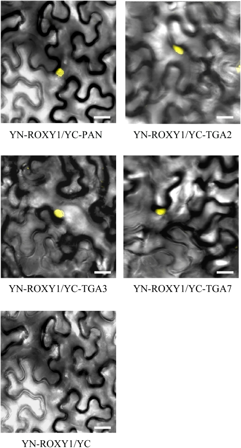
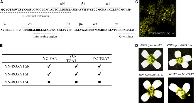
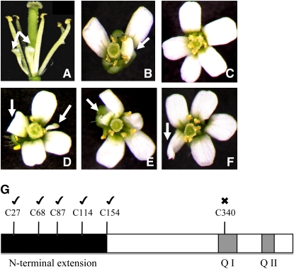
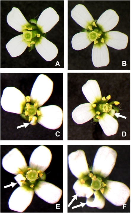
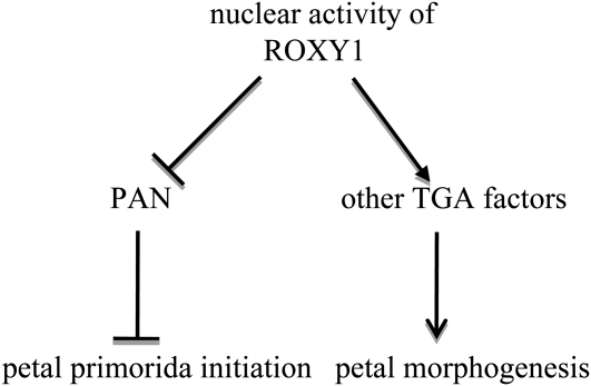
Glutaredoxins (GRXs) have thus far been associated mainly with redox-regulated processes participating in stress responses. However, ROXY1, encoding a GRX, has recently been shown to regulate petal primorida initiation and further petal morphogenesis in Arabidopsis thaliana. ROXY1 belongs to a land plant-specific class of GRXs that has a CC-type active site motif, which deviates from ubiquitously occurring CPYC and CGFS GRXs. Expression studies of yellow fluorescent protein-ROXY1 fusion genes driven by the cauliflower mosaic virus 35S promoter reveal a nucleocytoplasmic distribution of ROXY1. We demonstrate that nuclear localization of ROXY1 is indispensable and thus crucial for its activity in flower development. Yeast two-hybrid screens identified TGA transcription factors as interacting proteins, which was confirmed by bimolecular fluorescence complementation experiments showing their nuclear interaction in planta. Overlapping expression patterns of ROXY1 and TGA genes during flower development demonstrate that ROXY1/TGA protein interactions can occur in vivo and support their biological relevance in petal development. Deletion analysis of ROXY1 demonstrates the importance of the C terminus for its functionality and for mediating ROXY1/TGA protein interactions. Phenotypic analysis of the roxy1-2 pan double mutant and an engineered chimeric repressor mutant from PERIANTHIA (PAN), a floral TGA gene, supports a dual role of ROXY1 in petal development. Together, our results show that the ROXY1 protein functions in the nucleus, likely by modifying PAN posttranslationally and thereby regulating its activity in petal primordia initiation. Additionally, ROXY1 affects later petal morphogenesis, probably by modulating other TGA factors that might act redundantly during differentiation of second whorl organs.


Figures

Figure 1.
Figure 1.
Nuclear Localization of ROXY1 Is Required for Normal Petal Development. Fusion proteins of ROXY1 engineered by N-terminally incorporating an NLS and/or three YFP fragments were used to examine contributions of cytoplasmic versus nuclear ROXY1 localization to petal development. (A) Subcellular localization of ROXY1 fusion proteins transiently expressed in N. benthamiana leaves. Fluorescent images show different intracellular localizations of genetically engineered ROXY1 fusion proteins. Bars = 50 μm. (B) Complementation analysis of ROXY1 fusion genes. Indicated ROXY1 fusion genes driven under the control of the CaMV 35S promoter were transformed in roxy1-2 mutants. At least 58 transgenic T1 plants were examined for each construct, and representative pictures of petal phenotypes are shown.
Figure 2.
Figure 2.
ROXY1 Interacts with TGA Factors in the Nuclei of Transiently Transformed N. benthamiana Leaves. The BiFC technique was employed to investigate ROXY1 interactions with TGA factors in planta. The N terminus of YFP (YN) was cloned in-frame upstream of ROXY1. The C terminus of YFP (YC) was cloned in-frame upstream of TGA2, TGA3, TGA7, and PAN individually. Constructs were pairwise transiently expressed in N. benthamiana leaves. Reconstitution of YFP fluorescence was examined by confocal microscopy 3 to 5 d after transient coexpression of protein pairs, and representative pictures are shown. Yellow fluorescence in the nucleus was detected for interactions of ROXY1 with PAN, TGA2, TGA3, and TGA7, respectively. As a negative control, coexpression of YN-ROXY1 with nonfused YC failed to reconstitute a fluorescent YFP chromophore. Bars = 50 μm.
Figure 3.
Figure 3.
The C terminus of ROXY1 Is Crucial for Interaction with TGA Factors and Complementation of roxy1-2 Mutants. (A) The amino acid sequence of ROXY1 is shown using the one-letter code. The ROXY1-specific N-terminal extension, intervening region, and C terminus are highlighted with dashed lines below the sequence. Amino acid residues that form α-helices (αN, α1-3, and αC) and β-sheets (β1-4) are indicated by solid lines above the sequence. (B) Interactions between three mutated ROXY1 versions (ΔN1-38, ΔI85-98, and ΔC129-136) and PAN, TGA3, and TGA7 in the nuclei of transiently transformed N. benthamiana leaves using BiFC. Ticks indicate that YFP fluorescence was reconstituted by an interaction, whereas crosses denote the inability to detect an interaction. (C) Nucleocytoplasmic localization of the transiently expressed YFP-ROXY1ΔC fusion protein in N. benthamiana leaves was determined by confocal laser scanning microscopy, proving that the C-terminal deletion of ROXY1 does not affect its intracellular localization. Bar = 50 μm. (D) Complementation analysis of the three mutant versions of ROXY1. Wild-type and the three mutagenized ROXY1 genes driven by endogenous ROXY1 regulatory sequences were transformed into roxy1-2 mutant plants. Petal phenotypes of transgenic T1 plants were examined, and representative phenotypes are shown.
Figure 4.
Figure 4.
Comparisons of the C termini for Arabidopsis CC-Type GRXs. (A) Phylogenetic tree of Arabidopsis CC-type GRXs. Numbers at the branches indicate the bootstrap values for 1000 replicates. The alignment used to generate this tree is available as Supplemental Data Set 1 online. Except for the characterized ROXY1/2, all other CC-type GRXs were named successively as ROXY3 to ROXY21, according to decreasing protein sequence similarities with ROXY1. Respective Arabidopsis Genome Initiative codes and active site motifs are indicated, and the previously described GRX480 is denoted. Ticks indicate CC-type GRXs that were able to complement roxy1-2 mutants, and crosses denote those that did not exhibit a ROXY1-like activity and failed to complement the mutant. Expression of CC-type GRXs was driven under the control of ROXY1 regulatory sequences. Unmarked ROXYs were not tested in this study. (B) Alignment of C-terminal extensions of CC-type GRXs chosen for complementation experiments. Bold letters indicate a conserved Gly, representing the putative glutathione binding sites (G110 in ROXY1). The C-terminal four amino acids A(L/I)WL indicated by a gray box are conserved for all tested CC-type GRXs that could rescue the roxy1-2 mutant petal phenotype.
Figure 5.
Figure 5.
Overlapping Expression Domains of ROXY1, PAN, and TGA2 in Floral Tissues. ROXY1, PAN, and TGA2 antisense probes were hybridized to longitudinal sections through wild-type Arabidopsis flowers. c, carpel; fl, young flower; st, stamen; pe, petal. Bars = 50 μm. (A) ROXY1 is expressed (blue color) in the inflorescence apex at positions where future floral primordia will be formed. Expression in young flower buds indicates the initiation of sepal and stamen primordia. (B) and (C) PAN (B) and TGA2 (C) expression in young flowers is less distinct than that of ROXY1. PAN mRNA is detected in several cell layers of the inflorescence apical meristem and the upper half of a young flower bud. Likewise, TGA2 is expressed in cells of the upper floral dome. (D) to (F) In older flowers, after the initiation of floral organs, ROXY1 shows a distinct expression in young petal and stamen primordia (D), whereas PAN (E) and TGA2 (F) expression patterns are broader and their mRNAs are located in all cells of developing floral organs. (G) to (I) Later in flower development, ROXY1 (G), PAN (H), and TGA2 (I) mRNAs are localized to the inner carpel surface and delineate the area where ovule primordia will be initiated. Asterisks indicate inflorescence apices in (A) and (B). The arrow in (G) points to the ROXY1 expression domain.
Figure 6.
Figure 6.
Interaction between ROXY1 and PAN. (A) and (B) show typical petal phenotypes of roxy1-2 mutants, where petals remain often smaller or folded. The roxy1-2 pan double mutant (D) to (F) develops pentamerous flowers resembling those of the pan single mutant (C). Additionally, later petal morphogenesis is affected in double mutants that also form smaller (D) or folded petals in (D) to (F), thus resembling later roxy1-2 petal differentiation. (G) C340 is essential for PAN functionality during flower development. Schematic representation of the PAN protein indicates the positions of its six Cys residues (top), a specific N-terminal extension (black box), and two conserved Gln-rich regions (Q I and Q II, gray boxes). C27, C68, C87, C114, and C154 and C340 were individually mutagenized into Ser residues. For complementation experiments, the pan mutant plants were transformed with wild-type and mutated versions of PAN genes driven under the control of the CaMV 35S promoter. Flower phenotypes of transgenic T1 plants carrying each construct were examined. Ticks or crosses above Cys residues denote if mutagenized PAN proteins were still able to rescue the pan mutant phenotype (tick) or failed to exhibit a wild type-like PAN protein activity (cross). Arrows indicate altered petal morphogenesis, such as smaller or folded petals.
Figure 7.
Figure 7.
Petal Phenotypes Induced by the Chimeric PAN Repressor. To overcome functional redundancy of TGA factors, PAN was fused to the EAR-motif repression domain (SRDX). Ectopic expression of PANSRDX induced severe defects during flower development (500 flowers, 10 flowers from 50 T1 plants, were examined). Floral phenotypes of 35Spro:PANSRDX T1 transgenic plants grouping into four distinct classes are shown. (A) Tetramerous wild-type-like flowers with four normal petals (10.6%). (B) Pentamerous flowers with five normal petals resembling those of the single pan mutant (19.0%). (C) and (D) Tetramerous flowers with normal, smaller, or folded petals resembled those of the single roxy1-2 mutant (19.6%). The majority of analyzed flowers was pentamerous (50.8%) and developed normal but also smaller (E) or folded petals (F), thus resembling those of the roxy1-2 pan double mutant. Abnormal petals, such as smaller or folded ones, are marked by arrows.
Figure 8.
Figure 8.
ROXY1 Activities during Petal Development. Intracellular localization studies coupled with complementation data prove that the nuclear activity of ROXY1 is required for petal development. Genetic and protein interaction studies unraveled two distinct ROXY1 functions during petal organogenesis. At the early stage of petal development, ROXY1 regulates the number of petal primordia probably by negatively regulating the PAN activity. PAN suppresses a pentamerous flower patterning and reduces the petal organ number such that a tetramerous second whorl is formed. Later in petal development, ROXY1 likely positively regulates other TGA factors, which seem to act redundantly to govern normal petal morphogenesis. Arrows and hatchets indicate positive and negative interactions, respectively.

References

    1. Bandyopadhyay, S., Gama, F., Molina-Navarro, M.M., Gualberto, J.M., Claxton, R., Naik, S.G., Huynh, B.H., Herrero, E., Jacquot, J.P., Johnson, M.K., and Rouhier, N. (2008). Chloroplast monothiol glutaredoxins as scaffold proteins for the assembly and delivery of [2Fe-2S] clusters. EMBO J. 27 1122–1133. - PMC - PubMed
    1. Buchanan, B.B., and Balmer, Y. (2005). Redox regulation: A broadening horizon. Annu. Rev. Plant Biol. 56 187–220. - PubMed
    1. Cheng, N.H., Liu, J.Z., Brock, A., Nelson, R.S., and Hirschi, K.D. (2006). AtGRXcp, an Arabidopsis chloroplastic glutaredoxin, is critical for protection against protein oxidative damage. J. Biol. Chem. 281 26280–26288. - PubMed
    1. Chuang, C.F., Running, M.P., Williams, R.W., and Meyerowitz, E.M. (1999). The PERIANTHIA gene encodes a bZIP protein involved in the determination of floral organ number in Arabidopsis thaliana. Genes Dev. 13 334–344. - PMC - PubMed
    1. Clough, S.J., and Bent, A.F. (1998). Floral dip: A simplified method for Agrobacterium-mediated transformation of Arabidopsis thaliana. Plant J. 16 735–743. - PubMed

Publication types

MeSH terms

Associated data

LinkOut - more resources