. 2009 Apr;8(4):560-72.
doi: 10.1128/EC.00111-08. Epub 2009 Feb 13.

p21-activated kinases Cla4 and Ste20 regulate vacuole inheritance in Saccharomyces cerevisiae

Affiliations

p21-activated kinases Cla4 and Ste20 regulate vacuole inheritance in Saccharomyces cerevisiae

Clinton R Bartholomew et al. Eukaryot Cell. 2009 Apr.

Abstract

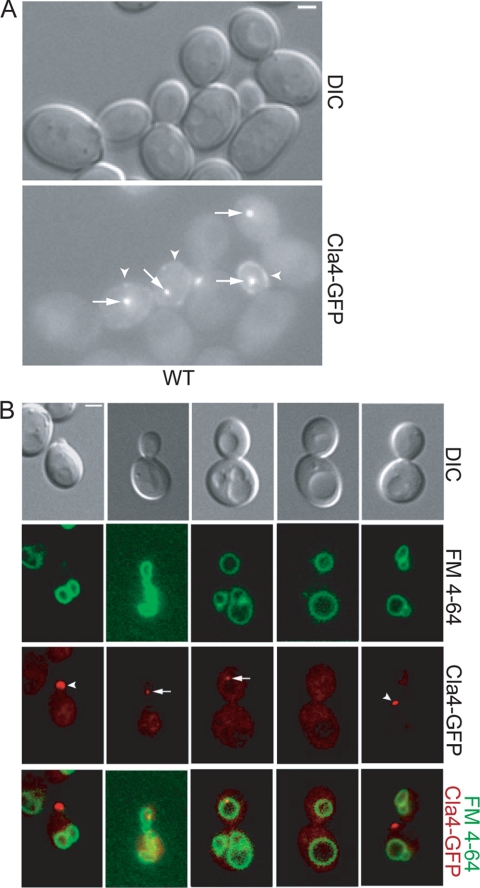
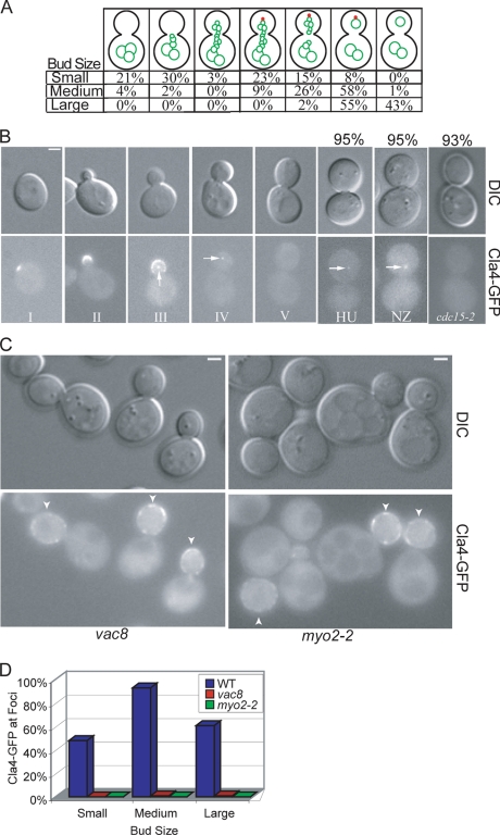
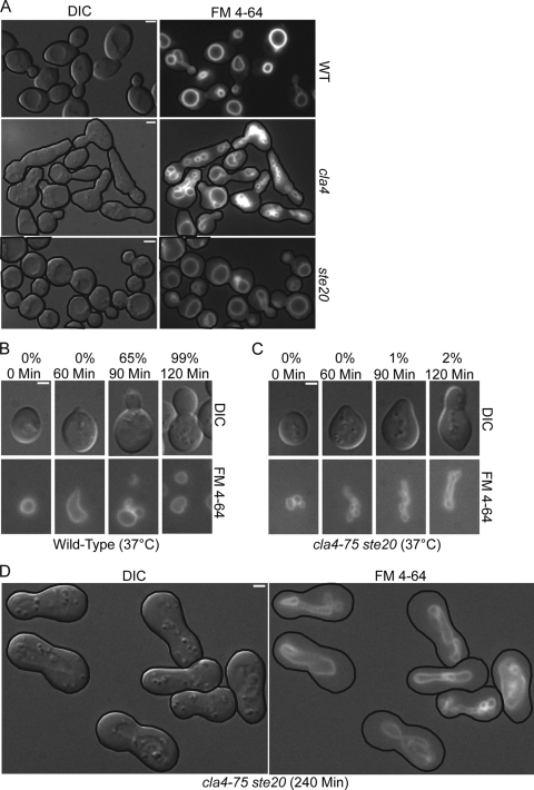
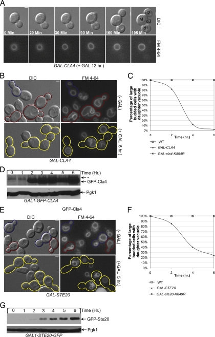
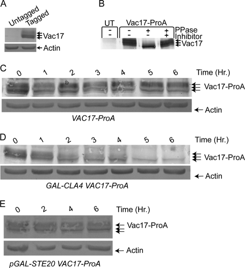
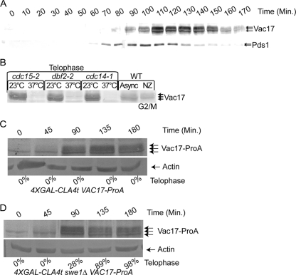
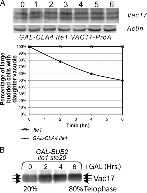
Each time Saccharomyces cerevisiae cells divide they ensure that both the mother and daughter cell inherit a vacuole by actively transporting a portion of the vacuole into the bud. As the mother cell begins budding, a tubular and vesicular segregation structure forms that is transported into the bud by the myosin V motor Myo2, which is bound to the vacuole-specific myosin receptor, Vac17 (41, 59, 70, 79). Upon arriving in the bud the segregation structure is resolved to found the daughter vacuole. The mechanism that regulates segregation structure resolution in a spatially dependent manner is unknown. In addition to resolving the segregation structure, Vac17 is degraded specifically in the bud to provide directionality to vacuole inheritance. It has been proposed that bud-specific degradation of Vac17 is promoted by proteins localized to or activated solely in the bud (77). The p21-activated kinases (PAKs) Cla4 and Ste20 are localized to and activated in the bud. Here we report that Cla4 is localized to the segregation structure just prior to segregation structure resolution, and cells lacking PAK function fail to resolve the segregation structure. Overexpression of either Cla4 or Ste20 inhibited vacuole inheritance and this inhibition was suppressed by the expression of nondegradable VAC17. Finally, PAK activity was required for Vac17 degradation in late M phase and CLA4 overexpression promoted Vac17 degradation. We propose that Cla4 and Ste20 are bud-specific proteins that play roles in both segregation structure resolution and the degradation of Vac17.


Figures

FIG. 1.
FIG. 1.
Cla4 localizes with the vacuole. (A) Cla4-GFP was visualized in asynchronous wild-type (YCH3250) cells. Arrows indicate perivacuolar Cla4-GFP localization, and arrowheads indicate cortex localization. (B) CLA4-GFP (YCH3250) cells were FM 4-64 stained and examined by fluorescence microscopy.
FIG. 2.
FIG. 2.
Cla4 requires vacuole inheritance for vacuole-associated punctate structure localization and remains on the daughter vacuole until late M phase. (A) Asynchronous cultures of FM 4-64-stained Cla4-GFP (YCH3250) were examined by fluorescence microscopy and each cell was categorized as small, medium, or large budded (>200 cells were counted for each bud size). Each cell was examined for both vacuole morphology (green) and Cla4-GFP localization to a punctate spot (red), and all cells fell into one of the categories shown. The percentages of small, medium, or large budded cells with the indicated vacuole morphology and punctate Cla4 localization are listed. (B) CLA4-GFP (YCH3250) localization was visualized in cells of various bud sizes and in cells arrested in S phase with hydroxyurea (HU), at G2/M with nocodazole (NZ), or at mitotic exit by arresting cdc15-2 (YCH1028). Values indicate the percentages of cells with the displayed Cla4-GFP localization. (C) Cla4-GFP was visualized in vac8 (YCH1665) and myo2-2 (YCH1752) cells. Arrowheads indicate Cla4-GFP localization at the cortex. (D) Cla4-GFP and FM 4-64-stained vacuole localization was quantified in small, medium, and large budded wild-type (YCH3250), vac8 (YCH1665), and myo2-2 (YCH1752) cells (>200 cells were counted for each bud size).
FIG. 3.
FIG. 3.
Cells lacking PAK function form but do not resolve segregation structures. (A) Mid-logarithmic-phase FM 4-64-strained wild-type (W303-1A), cla4 (KN3591), and ste20 (KN3621) strains were examined by fluorescence microscopy. (B to D) Asynchronous FM 4-64-stained wild-type (W303-1A) and cla4-75 ste20 (KN4580) cells were elutriated to obtain a uniform population of G1 cells, grown at 37°C, and examined at the indicated times by fluorescence microscopy. Values in panels B and C are the percentages of cells with a daughter vacuole.
FIG. 4.
FIG. 4.
Cells overexpressing CLA4 or STE20 have a vacuole inheritance defect. (A) Video microscopy and pedigree analysis of FM 4-64-stained GAL-CLA4 (YMG694) cells was performed on cells grown in YP plus 2% galactose. (B) FM 4-64-stained GAL-CLA4 (YMG694) cells were grown in YP plus 2% raffinose to mid-logarithmic stage. At time zero 2% galactose was added to half the culture and the presence of segregation structures in small and medium budded cells and of daughter vacuoles in large budded cells was examined. Representative pictures at the 6-h time point are shown. (C) Wild-type (W303-1A), GAL-CLA4 (YMG694), and GAL-cla4-K594R (YCB91) cells were treated as for panel B, and the percentage of large budded cells with a discrete daughter vacuole was quantified by fluorescence microscopy (>200 large budded cells were counted for each strain). (D) Mid-logarithmic-phase GAL-GFP-CLA4 (YCH5295) cells were grown in YP plus 2% raffinose. At the zero time point 2% galactose was added to induce GFP-CLA4 overexpression, samples were taken every hour, and Western blot analysis was performed. (E and F) FM 4-64-stained pRS316 (YCH4811), p316-GAL-GST-STE20 (YCH4981), and pRS316-GAL-GST-ste20-K649R (YCH4982) cells were grown in SC-URA plus 2% raffinose to the mid-logarithmic stage and treated as for panels B and C. Representative pictures at the 0- and 5-h time points are shown for GAL-STE20 cells. (G) Mid-logarithmic-phase GAL-STE20-GFP (YCB94) cells were treated as for panel D. Pedigree analysis: M, mother; d1 to -3, primary, secondary, and tertiary daughters.
FIG. 5.
FIG. 5.
Vac17 is a phosphoprotein and CLA4-overexpressing cells exhibit a decrease in Vac17 levels. (A) Vac17-ProA and actin protein levels were examined by Western blot analysis in untagged (W303-1A) and VAC17-ProA (YCH4774) cells. (B) Vac17-ProA was immunoprecipitated from protein extracts of 4X GAL-CLA4t swe1Δ VAC17-ProA (YCH4893) or untagged (UT) 4X GAL-CLA4t swe1 (Piatti2711) strains grown on YP plus 2% galactose medium for 6 h. Immunoprecipitated material was split and treated with buffer alone, λ-phosphatase, or λ-phosphatase plus phosphatase inhibitors. (C and D) Mid-logarithmic-phase VAC17-ProA (YCH4774) and GAL-CLA4 VAC17-ProA (YCH4882) cells were grown in YP plus 2% raffinose. At the zero time point 2% galactose was added to induce CLA4 overexpression, samples were taken every hour, and Western blot analysis was performed. (E) Mid-logarithmic-phase GAL-STE20 VAC17-ProA (YCH4843) cells were grown in SC-TRP plus 2% raffinose. At the zero time point 2% galactose was added to induce STE20 overexpression, samples were taken every hour, and Western blot analysis was performed.
FIG. 6.
FIG. 6.
PAK function is required for Vac17-ProA degradation in late M phase. (A) VAC17-ProA PDS1-HA (YCH5162) cells were arrested in G1 with α-factor and released, samples were taken every 10 min, and Vac17-ProA and Pds1-HA were examined by Western blot analysis. (B) Vac17-ProA levels were assayed by Western blotting in cdc15-2 (YCH4862), dbf2-2 (YCH4869), and cdc14-1 (YCH4852) cells grown at 23°C or arrested at telophase in 37°C medium. (C and D) 4XGAL-CLA4t VAC17-ProA (YCH4894) and 4XGAL-CLA4t swe1 VAC17-ProA (YCH4893) cells were α-factor arrested and released, and samples were taken at the indicated times and subjected to Western blot analysis. Additionally, the percentage of telophase cells was determined by DAPI staining and the number of cells with mother and bud nuclei staining was scored.
FIG. 7.
FIG. 7.
LTE1 is required for degradation of Vac17 in CLA4-overexpressing cells. (A) Mid-logarithmic-phase lte1 (YCH4737) and GAL-CLA4 lte1 VAC17-ProA (YCH4934) cells were grown in YP plus 2% raffinose and FM 4-64 stained. At the zero time point 2% galactose was added to induce CLA4 overexpression, samples were taken every hour, and Western blot analysis was performed and vacuole inheritance was quantified. (B) Mid-logarithmic-phase GAL-BUB2 bub2 lte1 ste20 Vac17-ProA (YCH5286) cells were grown in YP plus 2% raffinose, 2% glucose was added at time zero, and samples were taken at the indicated times and subjected to Western blot analysis. Additionally, the percentage of telophase cells was determined by DAPI staining and scoring the number of cells with mother and bud nuclei staining.

References

    1. Aitchison, J. D., M. P. Rout, M. Marelli, G. Blobel, and R. W. Wozniak. 1995. Two novel related yeast nucleoporins Nup170p and Nup157p: complementation with the vertebrate homologue Nup155p and functional interactions with the yeast nuclear pore-membrane protein Pom152p. J. Cell Biol. 1311133-1148. - PMC - PubMed
    1. Ash, J., C. Wu, R. Larocque, M. Jamal, W. Stevens, M. Osborne, D. Y. Thomas, and M. Whiteway. 2003. Genetic analysis of the interface between Cdc42p and the CRIB domain of Ste20p in Saccharomyces cerevisiae. Genetics 1639-20. - PMC - PubMed
    1. Barr, F. A. 2002. Inheritance of the endoplasmic reticulum and Golgi apparatus. Curr. Opin. Cell Biol. 14496-499. - PubMed
    1. Benton, B. K., A. Tinkelenberg, I. Gonzalez, and F. R. Cross. 1997. Cla4p, a Saccharomyces cerevisiae Cdc42p-activated kinase involved in cytokinesis, is activated at mitosis. Mol. Cell. Biol. 175067-5076. - PMC - PubMed
    1. Bokoch, G. M. 2003. Biology of the p21-activated kinases. Annu. Rev. Biochem. 72743-781. - PubMed

MeSH terms

Substances