. 2009 Mar 3;106(9):3372-7.
doi: 10.1073/pnas.0813306106. Epub 2009 Feb 13.

Role of COX-2 in epithelial-stromal cell interactions and progression of ductal carcinoma in situ of the breast

Affiliations

Role of COX-2 in epithelial-stromal cell interactions and progression of ductal carcinoma in situ of the breast

Min Hu et al. Proc Natl Acad Sci U S A. .

Abstract

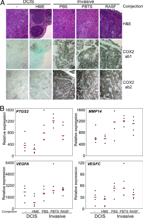
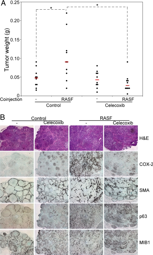
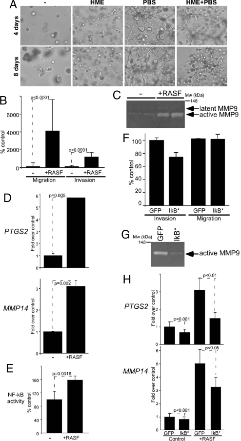
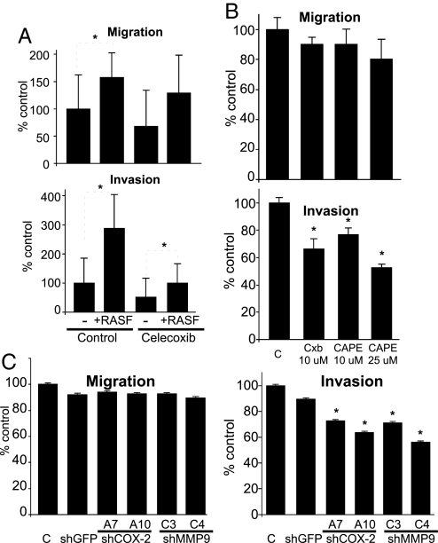
Epithelial-stromal cell interactions have an important role in breast tumor progression, but the molecular mechanisms underlying these effects are just beginning to be understood. We previously described that fibroblasts promote, whereas normal myoepithelial cells inhibit, the progression of ductal carcinoma in situ (DCIS) to invasive breast carcinomas by using a xenograft model of human DCIS. Here, we report that the tumor growth and progression-promoting effects of fibroblasts are at least in part due to increased COX-2 expression in tumor epithelial cells provoked by their interaction with fibroblasts. Up-regulation of COX-2 in DCIS xenografts resulted in increased VEGF and MMP14 expression, which may contribute to the larger weight and invasive histology of COX-2-expressing tumors. Administration of celecoxib, a selective COX-2 inhibitor, to tumor-bearing mice decreased xenograft tumor weight and inhibited progression to invasion. Coculture of fibroblasts with DCIS epithelial cells enhanced their motility and invasion, and this change was associated with increased MMP14 expression and MMP9 protease activity. We identified the NF-kappaB pathway as one of the mediators of stromal fibroblast-derived signals regulating COX-2 expression in tumor epithelial cells. Inhibition of NF-kappaB and COX-2 activity and down-regulation of MMP9 expression attenuated the invasion-promoting effects of fibroblasts. These findings support a role for COX-2 in promoting the progression of DCIS to invasive breast carcinomas, and suggest that therapeutic targeting of the NF-kappaB and prostaglandin signaling pathways might be used for the treatment and prevention of breast cancer.


Conflict of interest statement

Conflict of interest statement: K.P. receives research support from and is a consultant to Novartis Pharmaceuticals, Inc., and is also a consultant to Aveo Pharmaceuticals, Inc., and Genego Inc.

Figures

Fig. 1.
Fig. 1.
The expression of COX-2 and angiogenic factors in MCFDCIS xenografts. (A) Immunohistochemical analysis of COX-2 expression in MCFDCIS xenografts using an antibody specific for human COX-2 (ab1) and one that also recognizes murine COX-2 (ab2). Low expression was detected in MCFDCIS cells injected alone (−) or coinjected with HME cells, whereas coinjection of any fibroblast (e.g., PBS, PBTS, and RASF) up-regulated COX-2 expression in tumor epithelial cells. No COX-2-positive cells were detected in the stroma. (B) Quantitative RT-PCR analysis of human-specific COX-2 (PTGS2), MMP14, VEGFA, and VEGFC gene expression in MCFDCIS xenografts. Decreased and increased expression of these genes was detected in MCFDCIS cells coinjected with HME cells and fibroblasts (PBS, PBTS, and RASF), respectively, relative to human-specific hypoxanthine phosphoribosyltransferase 1 (HPRT1) used as loading control.
Fig. 2.
Fig. 2.
The effect of celecoxib on MCFDCIS xenografts. The effect of a selective COX-2 inhibitor (celecoxib) on the weight (A) and histology (B) of MCFDCIS xenografts derived from cells injected alone (−) or coinjected with RASFs on control or celecoxib-containing diet. Xenografts from MCFDCIS cells alone (−) had DCIS histology and low COX-2 expression, regardless of diet. Tumors from MCFDCIS cells coinjected with RASF showed invasive phenotype and high COX-2 levels in the control diet group. Celecoxib abolished the tumor growth-stimulating effects of RASF (P = 0.0001), partially inhibited the progression to invasive tumors, and decreased COX-2 protein levels. No significant difference in the number of cycling (MIB1+) cells was detected after celecoxib treatment, but this observation does not exclude the possibility of decreased proliferation due to increased cell cycle length or increased apoptosis after celecoxib treatment. Statistically significant (P < 0.05) differences in tumor weight are marked with dashed lines and asterisks.
Fig. 3.
Fig. 3.
The effect of RASF coculture on MCFDCIS cells. (A) Morphology of MCFDCIS cells cultured alone (−) or together with the indicated cell types for 4 and 8 days after plating them into Matrigel. HME cells suppressed the growth of MCFDCIS cells and decreased the invasion-promoting effects of fibroblasts. (B) Coculture with RASFs statistically significantly (P < 0.0001) increased the migration and invasion of MCFDCIS cells. (C) MMP9 gelatinase activity detected in zymograms using conditioned media of MCFDCIS cells cultured alone or cocultured with RASFs. Latent and activate forms of MMP9 (determined based on molecular weight) are marked with arrows. Migration of the molecular weight marker is indicated. (D) Quantitative RT-PCR analysis of PTGS2 (COX-2) and MMP14 expression in MCFDCIS cells cultured alone (−) or with RASFs (+RASF); y axis indicates relative mRNA levels normalized to RPL39. (E) Activation of NF-κB in MCFDCIS cells by RASF coculture; y axis indicates NF-κB dependent firefly luciferase activity relative to Renilla luciferase. (F) The expression of IκB* in MCFDCIS cells attenuated their enhanced invasion (P = 0.049) induced by RASF cells without significant effect on their motility (P = 0.949). (G) The activity of MMP9 induced by RASF coculture was reduced by expressing IκB* in MCFDCIS cells. Cells infected with GFP expressing adenovirus were used as controls. (H) IκB* expression in MCFDCIS cells attenuated the increase in PTGS2 and MMP14 mRNA levels induced by RASF coculture. Infection with GFP expressing adenovirus had no effect; y axis indicates mRNA levels normalized relative to that of RPL39.
Fig. 4.
Fig. 4.
Inhibition of COX-2 or NF-κB in cocultures. Inhibition of COX-2 activity by using celecoxib (A) or via inhibition of NF-κB activation using CAPE (B) inhibited the increased invasive capacity of MCFDCIS cells induced by RASF coculture, whereas cell motility was not affected. Relative migration and invasion compared with MCFDCIS cells cultured alone are indicated on the y axis. Statistically significant (P < 0.05) differences are marked with dashed lines and asterisks. (C) Down-regulation of COX-2 and MMP9 expression in MCFDCIS cells using lentiviral shRNA decreased their enhanced invasion, but not migration induced by RASF coculture; y axis indicates migration and invasion relative to MCFDCIS cells cultured alone.

References

    1. Nelson CM, Bissell MJ. Of extracellular matrix, scaffolds, and signaling: Tissue architecture regulates development, homeostasis, and cancer. Annu Rev Cell Dev Biol. 2006;22:287–309. - PMC - PubMed
    1. Weinberg R, Mihich E. Eighteenth annual Pezcoller symposium: Tumor microenvironment and heterotypic interactions. Cancer Res. 2006;66:11550–11553. - PubMed
    1. Tlsty TD, Coussens LM. Tumor stroma and regulation of cancer development. Annu Rev Pathol. 2006;1:119–150. - PubMed
    1. Hu M, Polyak K. Microenvironmental regulation of cancer development. Curr Opin Genet Dev. 2008;18:27–34. - PMC - PubMed
    1. Mani SA, et al. The epithelial-mesenchymal transition generates cells with properties of stem cells. Cell. 2008;133:704–715. - PMC - PubMed

Publication types

MeSH terms