. 2009 Mar 3;106(9):3154-9.
doi: 10.1073/pnas.0813110106. Epub 2009 Feb 13.

Interaction forces and adhesion of supported myelin lipid bilayers modulated by myelin basic protein

Affiliations

Interaction forces and adhesion of supported myelin lipid bilayers modulated by myelin basic protein

Younjin Min et al. Proc Natl Acad Sci U S A. .

Abstract

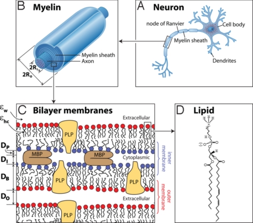
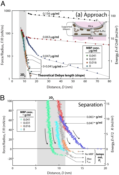
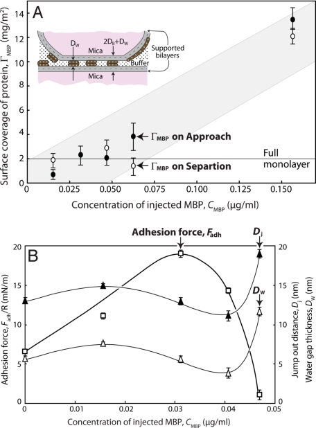
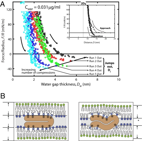
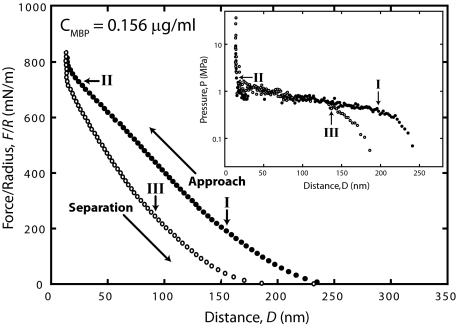
Force-distance measurements between supported lipid bilayers mimicking the cytoplasmic surface of myelin at various surface coverages of myelin basic protein (MBP) indicate that maximum adhesion and minimum cytoplasmic spacing occur when each negative lipid in the membrane can bind to a positive arginine or lysine group on MBP. At the optimal lipid/protein ratio, additional attractive forces are provided by hydrophobic, van der Waals, and weak dipolar interactions between zwitterionic groups on the lipids and MBP. When MBP is depleted, the adhesion decreases and the cytoplasmic space swells; when MBP is in excess, the bilayers swell even more. Excess MBP forms a weak gel between the surfaces, which collapses on compression. The organization and proper functioning of myelin can be understood in terms of physical noncovalent forces that are optimized at a particular combination of both the amounts of and ratio between the charged lipids and MBP. Thus loss of adhesion, possibly contributing to demyelination, can be brought about by either an excess or deficit of MBP or anionic lipids.


Conflict of interest statement

The authors declare no conflict of interest.

Figures

Fig. 1.
Fig. 1.
The structure of the myelin sheath. The myelinated axon (A), myelin sheath (B), bilayer membranes (C), and phosphatidylethanolamine (PE) (D) are shown. Each bilayer of thickness DB is separated by cytoplasmic and extracellular water gaps of thicknesses DI and DO and effective protein thickness DP occupied by the fraction of MBP constituting the cytoplasmic water gap.
Fig. 2.
Fig. 2.
Normalized force–distance profiles with different amounts of MBP in solution. F(D)/R measured on approach (A) and on separation (B) between 2 EAE cytoplasmic myelin bilayers in the presence of various amounts of MBP (in μg of injected MBP into the 80-mL incubating Mops buffer solution) at pH 7.2 and 24 °C. D = 0 corresponds to mica–mica contact, and DB represents 1 bilayer thickness as defined in Fig. 1. The right axis shows the corresponding interaction energy, E(D) = F(D)/2πR, per unit area between 2 planar bilayers, calculated according to the Derjaguin approximation (23). The equilibrium separation between planar bilayers corresponds to where F/R is a minimum; zero force in B is the equilibrium separation between 2 curved surfaces. Configuration of MBP in the bulk is primarily random coil (26, 37), which transforms into a C-shaped structure when it interacts with (bridges) 2 bilayers (38). Each curve corresponds to the second approach–separation cycle (compare Fig. 4). The numbers shown are the bulk MBP concentrations; the boxed concentrations refer only to the 4 curves in the concentration range 0–0.041 μg/mL, which have matching colored data points. The range of 2DB was obtained by subtracting the water layer thickness of Dw ≈ 2 nm from final “hard wall contact” distances.
Fig. 3.
Fig. 3.
Adsorbed density of MBP and adhesion forces with different amounts of MBP in solution. Calculations and measurements of injected MBP. (A) Calculated surface coverage of MBP (ΓMBP) adsorbed between and bridging 2 cytoplasmic EAE bilayers at increasing amounts, CMBP, of MBP injected into the 80-mL Mops buffer solution at pH 7.2 and 24 °C. Full monolayer coverage of MBP is shown by the line at 2 mg/m2 estimated from Eq. 6 by using φMBP = 1 and D = 3 nm, which corresponds to the thickness of C-shaped MBP (14). This coverage agrees with the measured ΓMBP at the maximum adhesion force and minimum water-gap thickness (see B). (B) Adhesion forces measured, corresponding jump-out distances [Dj (which equals 2DB + DI + DP)], and water-gap thicknesses [Dw (which equals DI + DP)] at CMBP ≤ 0.047 μg/mL]. Note that Fadh/R first increases and then decreases to zero after showing a maximum at ΓMBP ≈ 2 mg/m2, whereas Dj and DW pass through a minimum close to where the adhesion is maximum. Black and white symbols refer to values measured on approach and separation (shown in Fig. 3A). The Inset in A is a sketch of the adsorbed bilayers showing how MBP most likely acts to couple the 2 bilayers together. Note that the absorption or surface coverage in A may not be linear with the MBP concentration because MBP can grow as a gel layer on the surfaces (see Fig. 5). Most likely, the adsorption has 2 regimes, one to a full, close-packed MBP monolayer (as in A) and the other corresponding to the growth of a gel layer.
Fig. 4.
Fig. 4.
Effect of previous contacts and contact time on adhesion. (A) Adhesion forces measured on separation in Cmbp 0.031 μg/mL MBP solution under the same conditions as in Fig. 2B. Successive force runs show a progressively increasing adhesion and a concomitant decrease of the equilibrium (contact) separation brought about by a decreasing range of the stabilizing repulsive steric force. Water-gap thickness, DW, was obtained by subtracting 2DB ≈ 7.4 nm from the final contact separations. The contact time in each cycle was <1 min except in cycle 3, where the 2 surfaces were kept in contact for 30 min. Note the linear relationship between the adhesion forces, Fadh/R, and the jump-out distances, Dj, except for the noticeable deviation in run 3. Inset shows the total force cycle measured on approach and separation (compare Fig. 2) and the hysteresis in the forces (compare Fig. 5). A band of 2DB from 7.4–8.5 nm indicates a range of 2 bilayer thicknesses obtained by assuming a constant bilayer thickness and by subtracting a typical water-layer thickness (≈2 nm) from the final contact separation. (B) Possible model for the molecular rearrangements occurring on successive compressions that force the MBP molecules to penetrate deeper into the bilayers and enhance their hydrophobic contacts (15, 28). Note that MBP penetrates deeper into the bilayers with each successive compression. This mechanism would explain the decreasing range of the repulsion and the increasing adhesion. Note that DW* becomes less than DW after successive compressions whereas DB remains unchanged under the applied pressure range (<1 MPa).
Fig. 5.
Fig. 5.
Hysteretic forces in F/R vs. D runs measured in CMBP = 0.156 μg/ml MBP solution. (Inset) When the force F/R between the 2 curved surfaces is converted to the pressure P between 2 planar surfaces (using the Derjaguin approximation to obtain E, then differentiating to get P = dE/dD), and the calculated pressure vs. distance (proportional to the water gap volume) between 2 planar surfaces is plotted, the MBP behaves like a weak gel with a collapse pressure of P ≈ 1 MPa. The collapse resembles a first-order phase transition because it has constant (horizontal) pressure regime over a large range of DI (water volume) corresponding to a collapse to ≈10% of the original volume occupied. The arrows indicate where the slopes of F/R and pressure change, and the corresponding letters represent the sequences from I–III.

Similar articles

Cited by

References

    1. Raine CS. In: Myelin. Morell P, editor. New York: Plenum; 1984. pp. 1–41.
    1. Inouye H, Kirschner DA. Membrane interactions in nerve myelin. 1. Determination of surface-charge from effects of pH and ionic-strength on period. Biophys J. 1988;53:235–245. - PMC - PubMed
    1. Williams KA, Deber CM. The structure and function of central nervous system myelin. Crit Rev Clin Lab Sci. 1993;30:29–64. - PubMed
    1. Inouye H, Kirschner DA. Membrane interactions in nerve myelin. 2. Determination of surface-charge from biochemical data. Biophys J. 1988;53:247–260. - PMC - PubMed
    1. Jeans J. The Mathematical Theory of Electricity and Magnetism. Cambridge, UK: Cambridge Univ Press; 1966. pp. 332–335.

Publication types

LinkOut - more resources