. 2009 Mar 3;106(9):3615-20.
doi: 10.1073/pnas.0808717106. Epub 2009 Feb 13.

Control of Arabidopsis meristem development by thioredoxin-dependent regulation of intercellular transport

Affiliations

Control of Arabidopsis meristem development by thioredoxin-dependent regulation of intercellular transport

Yoselin Benitez-Alfonso et al. Proc Natl Acad Sci U S A. .

Abstract

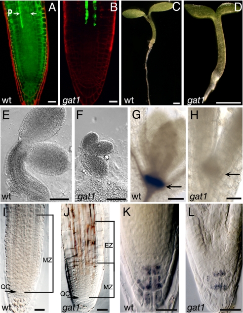
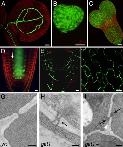
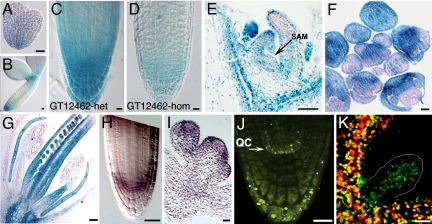
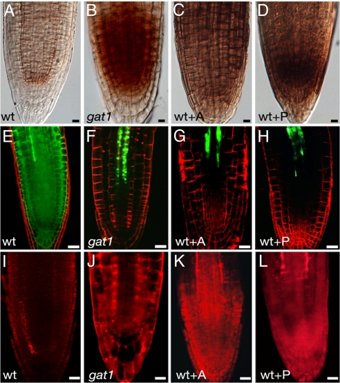
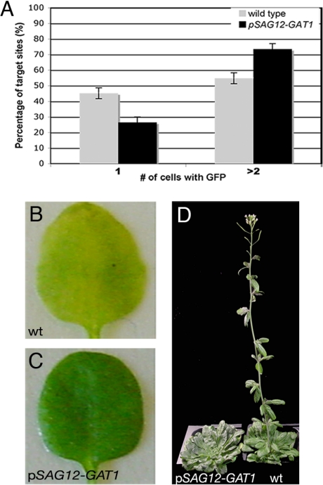
Cell-to-cell transport in plants occurs through cytoplasmic channels called "plasmodesmata" and is regulated by developmental and environmental factors. Callose deposition modulates plasmodesmal transport in vivo, but little is known about the mechanisms that regulate this process. Here we report a genetic approach to identify mutants affecting plasmodesmal transport. We isolated 5 mutants, named gfp arrested trafficking (gat), affected in GFP unloading from the phloem into the meristem. gat1 mutants were seedling lethal and carried lesions in an m-type thioredoxin that is expressed in non-green plastids of meristems and organ primordia. Callose and hydrogen peroxide accumulated in gat1 mutants, and WT plants subjected to oxidative conditions phenocopied the gat1 trafficking defects. Ectopic expression of GAT1 in mature leaves increased plasmodesmal permeability and led to a delay in senescence and flowering time. We propose a role for the GAT1 thioredoxin in the redox regulation of callose deposition and symplastic permeability that is essential for meristem maintenance in Arabidopsis.


Conflict of interest statement

The authors declare no conflict of interest.

Figures

Fig. 1.
Fig. 1.
GFP transport, embryo and seedling phenotypes of gat1. (A) WT seedlings expressing pSUC2-GFP show GFP diffusion out of the phloem (P, arrows) into the root meristem. (B) gat1 mutants show severe restriction in GFP transport out of the phloem. Seedling phenotypes of (C) WT and (D) gat1 mutants at 6 dpg show that gat1 is smaller than WT. (E, F) Embryos at cotyledon stage show that gat1 embryos also are smaller than WT. GUS staining of (G) WT and (H) gat1 siblings shows that a pSTM-GUS reporter is not expressed in the mutant (arrow indicates position of SAM). (I, J) The meristem zone (MZ) in gat1 roots at 6 dpg is smaller than in WT. The elongation zone (EZ) also is indicated in gat1 (J). Lugol's staining of (K) WT and (L) gat1 collumela cells shows that fewer starch granules accumulated in the mutant. Scale bars represent 20 μm in A, B, and I–L, 1 mm in C and D, 100 μm in E and F, and 50 μm in G and H.
Fig. 2.
Fig. 2.
gat1 PD phenotypes. gat1 seedlings expressing pSUC2-GFP, showing that GFP is restricted to the phloem in the shoot (A). Loading of HPTS in gat1 heart (B) and torpedo (C) embryo stages indicates symplastic transport. (D) gat1 seedlings expressing pSHR-SHR-GFP transport the fusion protein from the stele to the endodermis (white arrow). Expression of the fusion protein PDLP1A-YFP shows targeting to PDs in gat1 root (E) and cotyledons (F). TEM analysis of root sections at 50 μm from the QC shows simple PD in WT (G) but branched (H) and occluded PD (I) in gat1 (arrows). Scale bars represent 100 μm in A, 20 μm in B–D and F, 5 μm in E, and 0.2 μm in G–I.
Fig. 3.
Fig. 3.
GAT1 is expressed in meristems and primordia and localizes to plastids. GUS staining of the gene trap line gat1–3 shows expression in embryos from heart (A) and cotyledon (B) stages, in root meristem (C, D), vegetative apex including SAM vasculature (arrow) (E), inflorescences (F), and flowers (G). The homozygous gene trap insertion shows reduced expression (D) compared with heterozygote (C). In situ hybridization confirms GAT1 expression in root (H) and floral meristems (I). (J) Plants expressing GAT1:YFP under the endogenous promoter show native expression in non-green plastids of QC (arrows) and columella cells. (K) Confocal images of seedling shoots from plants overexpressing GAT1:YFP show green fluorescence in both photosynthetic and non-photosynthetic tissues (marked) that co-localizes with chlorophyll red autofluorescence. Scale bars represent 10 μm in A, B, and E and 20 μm in C, D, and F–K.
Fig. 4.
Fig. 4.
WT seedlings germinated in oxidants phenocopy gat1. DAB staining of (A) WT, (B) gat1, and WT seedlings treated with (C) alloxan (WT+A) or (D) paraquat (WT+P). gat1 and oxidant-treated seedlings accumulated ROS in the root meristem. WT seedlings expressing pSUC2-GFP and treated with (G) alloxan (WT+A) or (H) paraquat (WT+P) show defects in GFP diffusion and callose deposition (aniline blue staining shown in red, K, L) similar to those in gat1 mutants (F, J). Nontreated WT seedlings are shown in E and I. Scale bars represent 20 μm.
Fig. 5.
Fig. 5.
Ectopic expression of GAT1 promotes trafficking and delays senescence and flowering time. (A) The percentage of target sites in WT or pSAG12-GAT1 leaves that transported (> 2 cells) or did not transport (1 cell) GFP after bombardment with p35S-GFP. A higher percentage of events show GFP diffusion in pSAG12-GAT1. Dark-induced senescence of detached leaves from (B) WT and (C) pSAG12-GAT1 plants at 5 days after induction show that pSAG12-GAT1 leaves were delayed in senescence. pSAG12-GAT1 plants also were delayed in flowering in short-day conditions (D). Plants are 12 weeks old. The WT plant flowered 3 weeks earlier.

Comment in

References

    1. Benitez-Alfonso Y, Cantrill L, Jackson D. In: Cell-Cell Channels. Baluska F, Volkmann D, Barlow PW, editors. Austin, TX: Landes Bioscience; 2006.
    1. Crawford KM, Zambryski PC. Non-targeted and targeted protein movement through plasmodesmata in leaves in different developmental and physiological states. Plant Physiol. 2001;125:1802–1812. - PMC - PubMed
    1. Lucas WJ, Lee JY. Plasmodesmata as a supracellular control network in plants. Nat Rev Mol Cell Biol. 2004;5:712–726. - PubMed
    1. Cilia M, Jackson D. Plasmodesmata form and function. Curr Opin Cell Biol. 2004;16:500–506. - PubMed
    1. Kurata T, Okada K, Wada T. Intercellular movement of transcription factors. Curr Opin Plant Biol. 2005;8:600–605. - PubMed

Publication types

MeSH terms