. 2009 May;329(2):657-68.
doi: 10.1124/jpet.108.148007. Epub 2009 Feb 13.

Interferon-gamma released by gluten-stimulated celiac disease-specific intestinal T cells enhances the transepithelial flux of gluten peptides

Affiliations

Interferon-gamma released by gluten-stimulated celiac disease-specific intestinal T cells enhances the transepithelial flux of gluten peptides

Michael T Bethune et al. J Pharmacol Exp Ther. 2009 May.

Abstract

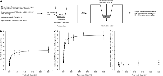
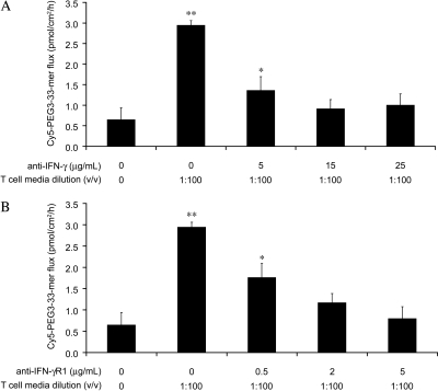
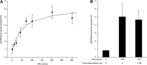
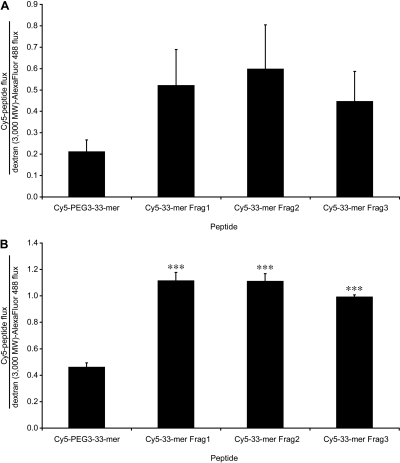
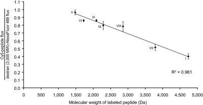
Celiac sprue is a T-cell-mediated enteropathy elicited in genetically susceptible individuals by dietary gluten proteins. To initiate and propagate inflammation, proteolytically resistant gluten peptides must be translocated across the small intestinal epithelium and presented to DQ2-restricted T cells, but the effectors enabling this translocation under normal and inflammatory conditions are not well understood. We demonstrate that a fluorescently labeled antigenic 33-mer gluten peptide is translocated intact across a T84 cultured epithelial cell monolayer and that preincubation of the monolayer with media from gluten-stimulated, celiac patient-derived intestinal T cells enhances the apical-to-basolateral flux of this peptide in a dose-dependent, saturable manner. The permeability-enhancing activity of activated T-cell media is inhibited by blocking antibodies against either interferon-gamma or its receptor and is recapitulated using recombinant interferon-gamma. At saturating levels of interferon-gamma, activated T-cell media does not further increase transepithelial peptide flux, indicating the primacy of interferon-gamma as an effector of increased epithelial permeability during inflammation. Reducing the assay temperature to 4 degrees C reverses the effect of interferon-gamma but does not reduce basal peptide flux occurring in the absence of interferon-gamma, suggesting active transcellular transport of intact peptides is increased during inflammation. A panel of disease-relevant gluten peptides exhibited an inverse correlation between size and transepithelial flux but no apparent sequence constraints. Anti-interferon-gamma therapy may mitigate the vicious cycle of gluten-induced interferon-gamma secretion and interferon-gamma-mediated enhancement of gluten peptide flux but is unlikely to prevent translocation of gluten peptides in the absence of inflammatory conditions.


Figures

Fig. 1.
Fig. 1.
Panel of synthetic gluten peptides labeled with Cy5 fluorescent dye. Structures are shown for the dye and PEG linker (where present). Sequences, common literature identifiers, and references containing original identification are provided for each peptide. In peptide I (Cy5-PEG3–33-mer), the 33-mer is linked via an amide bond to a PEG3 linker, which is linked to the dye. All other peptides used are directly linked to the dye via an amide bond.
Fig. 2.
Fig. 2.
Activated T-cell media enhances the apical-to-basolateral transepithelial flux of Cy5-PEG3–33-mer gluten peptide across a cultured T84 monolayer. A, experimental plan. Gluten was digested to peptide fragments and deamidated by treatment with transglutaminase 2. Deamidated gluten fragments were incubated overnight with DQ2+ antigen-presenting cells (9088), after which gluten-specific, DQ2-restricted T cells from celiac patient intestinal biopsies (P28 TCL1) were added. After 48 h, APCs and T cells were pelleted via centrifugation, and the supernatant was collected. This activated T-cell media were added at varied dilutions to the basolateral chamber of Transwell supports bearing cultured T84 epithelial cells. After 48 h of preincubation, the media in both the apical and basolateral chambers was replaced, and fluorescently labeled markers (3000 and 70,000 mol. wt. dextrans and gluten peptide, 33-mer) were added to the apical chamber at 2 μM each. Fluorescence in basolateral samples was measured over time to determine the apical-to-basolateral flux of each marker. Fluxes for Cy5-PEG3–33-mer (B), dextran (3000 mol. wt.)-Alexa Fluor-488 (C), and dextran (70,000 mol. wt.)-Texas Red 595 (D) are plotted against the dilution (v/v) of activated T-cell media used during preincubation. Mean ± S.D.s for triplicate wells are shown. Data are representative of more than three independent experiments.
Fig. 3.
Fig. 3.
The ability of activated T-cell media to enhance permeability is inhibited by blocking antibodies directed against either IFN-γ or IFN-γR1. Transwell supports bearing T84 epithelial cells were preincubated with 1:100 (v/v) basolateral activated T-cell media supplemented with varied concentrations of either anti-IFN-γ blocking antibody (A) or anti-IFN-γR1 blocking antibody (B). The translocation assay was performed as described in Fig. 2 to determine the apical-to-basolateral flux of Cy5-PEG3–33-mer (2 μM initial apical concentration). Mean ± S.D.s for triplicate wells are shown. Data are representative of more than three independent experiments. Statistical comparisons were performed with respect to the control lacking activated T-cell media. *, p < 0.05; **, p < 0.01.
Fig. 4.
Fig. 4.
Interferon-γ is the primary effector of transepithelial gluten translocation in activated T-cell media. Transwell supports bearing T84 epithelial cells were preincubated with varied concentrations of purified recombinant IFN-γ for 48 h (A) or with a saturating concentration (300 U/ml) of purified recombinant IFN-γ supplemented with 1:100 (v/v) activated T-cell media (B). After preincubation, a translocation assay was performed as described in Fig. 2 to determine the apical-to-basolateral flux of Cy5-PEG3–33-mer (2 μM initial apical concentration). Mean ± S.D.s for triplicate wells are shown. Data are representative of more than three independent experiments. Statistical comparisons were performed with respect to the control lacking IFN-γ or activated T-cell media. *, p < 0.05.
Fig. 5.
Fig. 5.
Gluten peptides are translocated intact across a cultured epithelial monolayer under normal and inflammatory conditions. Transwell supports bearing T84 epithelial cells were preincubated with media alone or with 600 U/ml IFN-γ in the basolateral chamber for 48 h. Peptide translocation assays were performed as described in Fig. 2, except with serum-free media and with an initial apical concentration of 20 μM Cy5-PEG3–33-mer. Samples were taken from the apical and basolateral chambers at 0 and 10 h for LC-MS analysis. Absorbance at 640 nm was monitored, and the mass spectra corresponding to A640 peaks were examined to confirm peptide identity. The peak eluting at a retention time of 9 to 10 min is intact Cy5-PEG3–33-mer. The peak eluting at a retention time of 8 min is Cy5-PEG3-LQ, signifying limited processing of the N terminus by T84 cells. A, apical media at 0 h, containing 20 μM Cy5-PEG3–33-mer. B, apical media after 10-h incubation in wells preincubated with 0 U/ml IFN-γ. C, apical media after 10-h incubation in wells preincubated with 600 U/ml IFN-γ. D, basolateral media at 0 h (=serum-free media). E, basolateral media after 10-h incubation in wells preincubated with 0 U/ml IFN-γ. F, basolateral media after 10-h incubation in wells preincubated with 600 U/ml IFN-γ. Data are representative of more than three similar experiments.
Fig. 6.
Fig. 6.
Gluten peptides are translocated across a cultured epithelial monolayer at sufficient levels to initiate and propagate inflammation. A, experimental plan. Transwell supports bearing T84 epithelial cells were preincubated with media alone or with 600 U/ml IFN-γ in the basolateral chamber for 48 h. Peptide translocation assays were performed as described in Fig. 2, except with 20 μM Cy5-PEG3–33-mer. Samples were taken from the basolateral chambers after 10 h and treated with vehicle or with 50 μg/ml TG2 for 2 h at 37°C in the presence of 5 mM CaCl2. Samples were boiled to quench TG2 activity, diluted 1:2 or 1:10 (v/v) into T-cell media containing DQ2+ 9088 APCs, and incubated overnight. The next day, gluten-specific, DQ2-restricted T cells were added to peptide-loaded APCs to a final composition of 187,500 cells each of 9088 APCs and T cells in a 250-μl volume. Cells were incubated for 48 h, after which they were pelleted via centrifugation, and the supernatant was collected. The concentration (units per milliliter) of IFN-γ secreted by T cells in response to basolateral media from Transwell supports preincubated with 0 U/ml IFN-γ (B) and 600 U/ml IFN-γ (C) was determined by a commercial sandwich ELISA, as described under Materials and Methods. Mean S.D.s for triplicate measurements are shown. Statistical comparisons were performed with respect to the corresponding controls in which no apical Cy5-PEG3–33mer was added. *, p < 0.05; ***, p < 0.001; ****, p < 0.0001.
Fig. 7.
Fig. 7.
Reducing the assay temperature to 4°C reverses the permeabilizing effect of IFN-γ but does not reduce the basal gluten peptide flux. Transwell supports bearing T84 epithelial cells were preincubated with media alone or with 300 U/ml IFN-γ in the basolateral chamber for 48 h at 37°C. Peptide translocation assays were performed as described in Fig. 2; however, some wells were assayed at 4°C rather than 37°C. Mean ± S.D.s for triplicate wells are shown. Data are representative of three similar experiments. Statistical comparisons were performed with respect to the controls lacking IFN-γ for each assay temperature. *, p < 0.05.
Fig. 8.
Fig. 8.
Gluten peptide translocation is not inhibited by MβCD. Transwell supports bearing T84 epithelial cells were preincubated with 300 U/ml IFN-γ in the basolateral compartment for 48 h. Peptide translocation assays were performed as described in Fig. 2, except either serum-free (black bars) or serum-containing (gray bars) media were used, and the endocytosis inhibitor MβCD was added at varied concentrations to the apical compartment with 2 μM each of fluorescent dextrans and Cy5-PEG3–33-mer. The apical-to-basolateral flux of Cy5-PEG3–33-mer in each well was normalized to the flux of dextran Alexa Fluor-488. Mean ± S.D.s for triplicate wells are shown.
Fig. 9.
Fig. 9.
Synthetic proteolytic fragments of the 33-mer gluten peptide exhibit increased transepithelial flux relative to intact 33-mer. Transwell supports bearing T84 epithelial cells were preincubated with media alone (A) or with 600 U/ml IFN-γ (B) in the basolateral chamber for 48 h. Peptide translocation assays were performed as described in Fig. 2 using either intact Cy5-PEG3–33-mer or Cy5-labeled synthetic 33-mer fragments corresponding to products of digestion of 33-mer by the glutenase EP-B2. The Cy5-peptide flux in each well is normalized to the corresponding dextran (3000 mol. wt.)-Alexa Fluor-488 flux. Mean ± S.D.s for triplicate wells are shown. Data are representative of two similar experiments. Statistical comparisons were performed with respect to the flux of full-length Cy5-PEG3–33-mer. ***, p < 0.001.
Fig. 10.
Fig. 10.
A panel of disease-relevant gluten peptides exhibits a linear and inverse correlation between transepithelial peptide flux and peptide molecular weight. Transwell supports bearing T84 epithelial cells were preincubated with 600 U/ml IFN-γ in the basolateral chamber for 48 h. Peptide translocation assays were performed as described in Fig. 2 using 20 μM Cy5-peptides numbered according to Fig. 1. I, Cy5-PEG3-LQLQPF(PQPQLPY)3PQPQLPF; III, Cy5-QLQPFPQPQLPY; V, Cy5-LPYPQPQ; VI, Cy5-LPYPQPQPF; VII, Cy5-FLQPQQPFPQQPQQPYPQQ PQQPFPQ; VIII, Cy5-LGQQQPFPPQQPYPQPQPF; IX, Cy5-VSFQQPQQQYPSSQ. The Cy5-peptide flux in each well is normalized to the corresponding dextran (3000 mol. wt.)-Alexa Fluor-488 flux. Mean ± S.D.s for triplicate wells are shown.

Similar articles

Cited by

References

    1. Alaedini A and Green PH (2005) Narrative review: celiac disease: understanding a complex autoimmune disorder. Ann Intern Med 142 289-298. - PubMed
    1. Arentz-Hansen H, Körner R, Molberg O, Quarsten H, Vader W, Kooy YM, Lundin KE, Koning F, Roepstorff P, Sollid LM, et al. (2000) The intestinal T cell response to alpha-gliadin in adult celiac disease is focused on a single deamidated glutamine targeted by tissue transglutaminase. J Exp Med 191 603-612. - PMC - PubMed
    1. Baert FJ, D'Haens GR, Peeters M, Hiele MI, Schaible TF, Shealy D, Geboes K, and Rutgeerts PJ (1999) Tumor necrosis factor alpha antibody (infliximab) therapy profoundly down-regulates the inflammation in Crohn's ileocolitis. Gastroenterology 116 22-28. - PubMed
    1. Bethune MT and Khosla C (2008) Parallels between pathogens and gluten peptides in celiac sprue. PLoS Pathog 4 e34. - PMC - PubMed
    1. Bethune MT, Ribka E, Khosla C, and Sestak K (2008) Transepithelial transport and enzymatic detoxification of gluten in gluten-sensitive rhesus macaques. PLoS ONE 3 e1857. - PMC - PubMed

Publication types

MeSH terms