. 2009 Apr;15(4):666-74.
doi: 10.1261/rna.1462109. Epub 2009 Feb 13.

Characterization of a mimivirus RNA cap guanine-N2 methyltransferase

Affiliations

Characterization of a mimivirus RNA cap guanine-N2 methyltransferase

Delphine Benarroch et al. RNA. 2009 Apr.

Abstract

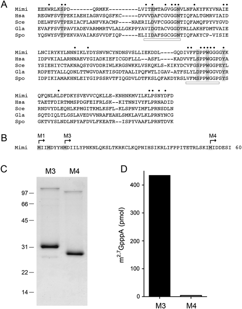
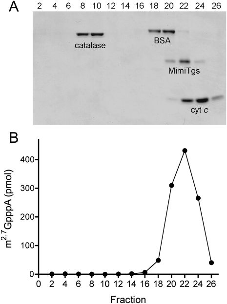
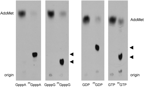
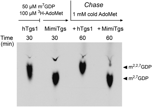
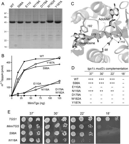
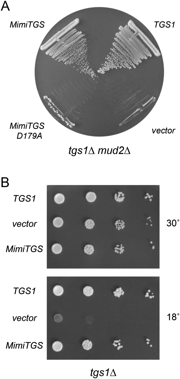
A 2,2,7-trimethylguanosine (TMG) cap is a signature feature of eukaryal snRNAs, telomerase RNAs, and trans-spliced nematode mRNAs. TMG and 2,7-dimethylguanosine (DMG) caps are also present on mRNAs of two species of alphaviruses (positive strand RNA viruses of the Togaviridae family). It is presently not known how viral mRNAs might acquire a hypermethylated cap. Mimivirus, a giant DNA virus that infects amoeba, encodes many putative enzymes and proteins implicated in RNA transactions, including the synthesis and capping of viral mRNAs and the promotion of cap-dependent translation. Here we report the identification, purification, and characterization of a mimivirus cap-specific guanine-N2 methyltransferase (MimiTgs), a monomeric enzyme that catalyzes a single round of methyl transfer from AdoMet to an m(7)G cap substrate to form a DMG cap product. MimiTgs, is apparently unable to convert a DMG cap to a TMG cap, and is thereby distinguished from the structurally homologous yeast and human Tgs1 enzymes. Nonetheless, we show genetically that MimiTgs is a true ortholog of Saccharomyces cerevisiae Tgs1. Our results hint that DMG caps can satisfy many of the functions of TMG caps in vivo. We speculate that DMG capping of mimivirus mRNAs might favor viral protein synthesis in the infected host.


Figures

FIGURE 1.
FIGURE 1.
Mimivirus Tgs. (A) The amino acid sequence of MimiTgs (Mimi) from amino acids 91–229 is aligned to the sequences of the homologous cap guanine-N2 methyltransferases of Homo sapiens Tgs1 (Hsa), Saccharomyces cerevisiae Tgs1 (Sce), Giardia lambia Tgs2 (Gla), and Schizosaccharomyces pombe Tgs1 (Spo). Gaps are indicated by dashes. (●) Positions of amino acid side-chain identity/similarity in all five proteins. The AdoMet-binding motif and a putative m7G-binding motif are denoted by horizontal bars. Positions in MimiTgs that were targeted for mutational analysis in the present study are highlighted in shaded boxes. (B) The N-terminal 60 amino acids of the predicted MimiTgs polypeptide are shown, with methionines highlighted in shaded boxes. The translation start sites for the recombinant MimiTgs proteins M1, M3, and M4 are indicated by arrows. (C) MimiTgs purification. Aliquots (5 μg) of the purified tag-free M3 and M4 proteins were analyzed by SDS-PAGE. The Coomassie Blue-stained gel is shown. The positions and sizes (in kDa) of marker polypeptides are indicated on the left. (D) Methyltransferase reaction mixtures (10 μL) containing 1 mM m7GpppA, 50 μM [3H-CH3]AdoMet, and 0.5 μg of purified M3 or M4 protein were incubated for 15 min at 37°C. The products were analyzed by ascending TLC in 0.05 M (NH4)2SO4. The extents of 3H-methyl transfer to the cap dinucleotide are shown.
FIGURE 2.
FIGURE 2.
Zonal velocity sedimentation. An aliquot (50 μg) of the tag-free wild-type MimiTgs protein was mixed with catalase (50 μg), BSA (50 μg), and cytochrome c (50 μg). The mixture was applied to a 5 mL 15%–30% glycerol gradient containing 50 mM Tris-HCl (pH 8.0), 500 mM NaCl, 2 mM DTT, 1 mM EDTA, 0.05% Triton X-100. The gradient was centrifuged at 50,000 rpm for 17 h at 4°C. Fractions (∼0.18 mL) were collected from the bottom of the tube. (A) Aliquots (20 μL) of the even-numbered gradient fractions were analyzed by SDS-PAGE. The Coomassie Blue-stained gel is shown, with the bottom (heaviest) gradient fraction at left and the top (lightest) fraction at right. The identities of the polypeptides are indicated. (B) Aliquots (1 μL) of the even-numbered fractions were assayed for cap guanine-N2 methyltransferase activity. The extents of methyl transfer from 50 μM [3H-CH3]AdoMet to 1 mM m7GpppA are plotted.
FIGURE 3.
FIGURE 3.
Methyl acceptor specificity of MimiTgs. Reaction mixtures (10 μL) containing 50 mM Tris-acetate (pH 6.0), 5 mM DTT, 50 μM [3H-CH3]AdoMet, 1 mM methyl acceptor as specified, and 1 μg MimiTgs were incubated for 60 min at 37°C. Aliquots (4 μL) were spotted on PEI-cellulose TLC plates, which were developed either with 0.05 M (NH4)2SO4 (for reactions with GpppA, m7GpppA, GpppG, and m7GpppG as methyl acceptors) or 0.1 M (NH4)2SO4 (for reactions with GDP and m7GDP as methyl acceptors) or 0.2 M (NH4)2SO4 (for reactions with GTP, and m7GTP as methyl acceptors). The chromatograms were treated with Enhance and 3H-labeled material was visualized by autoradiography. The methyltransferase reaction products m2,7GpppA, m2,7GpppG, m2,7GDP, and m2,7GTP are denoted by arrowheads at the right of the chromatograms.
FIGURE 4.
FIGURE 4.
MimiTgs catalyzes a single methyl addition at cap guanine-N2. Reaction mixtures containing 50 mM Tris-HCl (pH 8.0), 5 mM DTT, 100 μM [3H-CH3]AdoMet, 50 μM m7GDP, and either 1.8 μM purified recombinant human Tgs1(576–853) (hTgs1) or 0.8 μM MimiTgs were incubated for 30 min at 37°C. The MimiTgs reaction mixture was then split in half and supplemented with 1 mM “cold” AdoMet and either 7 μM hTgs1 or 7 μM MimiTgs, and incubation was continued for another 30 min. Aliquots were removed after the 30-min pulse-labeling phase and again after the cold AdoMet chase phase. The products were analyzed by PEI cellulose TLC in 0.1 M ammonium sulfate. The chromatogram was treated with Enhance and the 3H-labeled material was visualized by autoradiography.
FIGURE 5.
FIGURE 5.
Effects of alanine mutations on MimiTgs activity. (A) Aliquots (5 μg) of the nickel-agarose preparations of wild-type His10Smt3-MimiTgs and the indicated alanine mutants were analyzed by SDS-PAGE. Polypeptides were visualized by staining with Coomassie Blue dye. The positions and sizes (in kDa) of marker proteins are indicated on the left. (B) Reaction mixtures (10 μL) containing 50 mM Tris-acetate (pH 6.0), 5 mM DTT, 50 μM [3H-CH3]AdoMet, 1 mM m7GpppA, and proteins as specified were incubated for 15 min at 37°C. The extents of methyl transfer are plotted as a function of input enzyme. (C) Active site of Thermus thermophilus RNA guanine-N2 methyltransferase RsmC in complex with AdoMet and guanosine (from PDB 3DMH). Highlighted are the RsmC side chains that contact the methyl donor and acceptor and that have putative counterparts among the MimiTgs residues that were subjected to alanine scanning. Hydrogen bonding interactions are denoted by dashed lines. The residue numbers refer to the homologous positions in MimiTgs. (D,E) The ability of 2μ LEU2 plasmids bearing wild-type MimiTGS and the indicated alanine mutants to complement growth of a yeast tgs1Δ mud2Δ strain was tested by plasmid shuffle as described by Hausmann et al. (2008). Lethal mutations of MimiTGS were those that failed to support growth on medium containing 0.75 mg/mL 5-fluoroorotic acid (FOA) at any of the temperatures tested (scored as –). The tgs1Δ mud2Δ MimiTGS strains that grew on FOA at 30°C were tested for growth on rich medium by spotting serial 10-fold dilutions of liquid cultures (grown in SD-Leu medium) on YPD agar plates, which were then incubated at 18, 22, 30, or 37°C. Yeast tgs1Δ mud2Δ p(CEN LEU2 TGS1) cells were processed in parallel as a positive control. Photographs of the plates are shown in E. Growth of the viable MimiTGS-Ala strains was scored in D as follows: (+++) colony size indistinguishable from a strain bearing wild-type MimiTGS; (++) reduced colony size; (–) no growth.
FIGURE 6.
FIGURE 6.
MimiTgs is a functional ortholog of S. cerevisiae Tgs1. (A) Yeast tgs1Δ mud2Δ p (CEN URA3 TGS1) cells were transformed with a CEN LEU2 plasmid bearing wild-type S. cerevisiae TGS1 (positive control), an empty 2μ LEU2 vector (negative control), and 2μ LEU2 plasmids expressing either wild-type MimiTGS or MimiTGS-D179A. Leu+ transformants were selected at 30°C and then streaked to agar medium containing FOA. The plate was photographed after 3 d at 30°C. (B) Yeast tgs1Δ cells were transformed with a CEN LEU2 plasmid bearing wild-type S. cerevisiae TGS1 (positive control), an empty 2μ LEU2 vector (negative control), and a 2μ LEU2 plasmid expressing MimiTGS. Leu+ transformants were selected at 30°C and then tested for growth at 30°C and 18°C by spotting serial 10-fold dilutions of liquid cultures (grown at 30°C in SD-Leu medium) on Leu agar plates. The plates were photographed after incubation for 3 d at 30°C or 7 d at 18°C.

References

    1. Benarroch D., Smith P., Shuman S. Characterization of a trifunctional mimivirus mRNA capping enzyme and crystal structure of the RNA triphosphatase domain. Structure. 2008;16:501–512. - PubMed
    1. Busch H., Reddy R., Rothblum L., Choi Y.C. SnRNAs, SnRNPs, and RNA processing. Annu. Rev. Biochem. 1982;51:617–654. - PubMed
    1. Cai A., Jankowska-Anyszka M., Centers A., Chlebicka L., Stepinki J., Stolarski R., Darzynkiewicz E., Rhoads R.E. Quantitative assessment of mRNA cap analogues as inhibitors of in vitro translation. Biochemistry. 1999;38:8538–8547. - PubMed
    1. Claverie J.M., Ogata H., Audic S., Abergel C., Suhre K., Fournier P.E. Mimivirus and the emerging concept of “giant” virus. Virus Res. 2006;117:133–144. - PubMed
    1. Darzynkiewicz E., Stepinski J., Ekiel I., Haber D., Tahara S.M. β-globin mRNAs capped with m7G, m2,7G or m2,2,7G differ in intrinsic translation efficiency. Nucleic Acids Res. 1988;16:8953–8962. - PMC - PubMed

Publication types

LinkOut - more resources