. 2009 Apr 16;458(7240):899-903.
doi: 10.1038/nature07750.

Identification of a dendritic cell receptor that couples sensing of necrosis to immunity

Affiliations

Identification of a dendritic cell receptor that couples sensing of necrosis to immunity

David Sancho et al. Nature. .

Abstract

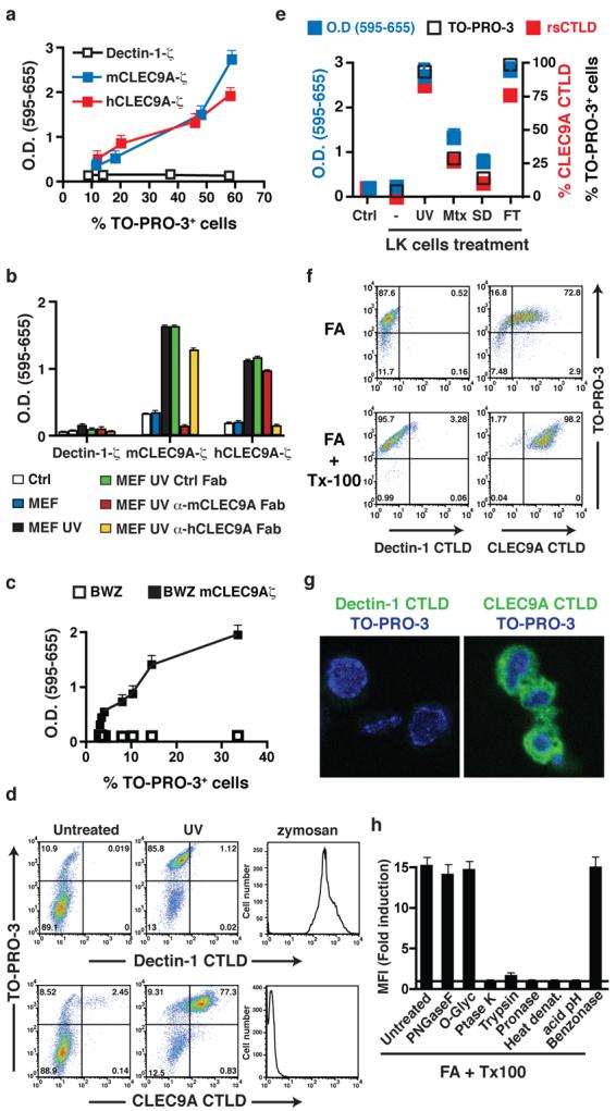
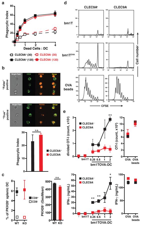
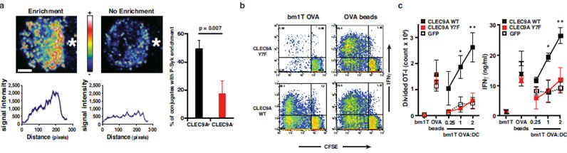
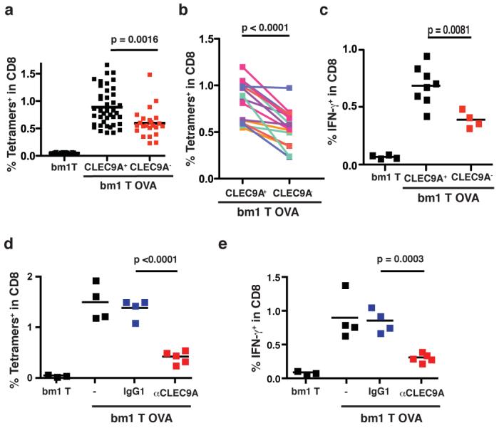
Injury or impaired clearance of apoptotic cells leads to the pathological accumulation of necrotic corpses, which induce an inflammatory response that initiates tissue repair. In addition, antigens present in necrotic cells can sometimes provoke a specific immune response and it has been argued that necrosis could explain adaptive immunity in seemingly infection-free situations, such as after allograft transplantation or in spontaneous and therapy-induced tumour rejection. In the mouse, the CD8alpha+ subset of dendritic cells phagocytoses dead cell remnants and cross-primes CD8+ T cells against cell-associated antigens. Here we show that CD8alpha+ dendritic cells use CLEC9A (also known as DNGR-1), a recently-characterized C-type lectin, to recognize a preformed signal that is exposed on necrotic cells. Loss or blockade of CLEC9A does not impair the uptake of necrotic cell material by CD8+ dendritic cells, but specifically reduces cross-presentation of dead-cell-associated antigens in vitro and decreases the immunogenicity of necrotic cells in vivo. The function of CLEC9A requires a key tyrosine residue in its intracellular tail that allows the recruitment and activation of the tyrosine kinase SYK, which is also essential for cross-presentation of dead-cell-associated antigens. Thus, CLEC9A functions as a SYK-coupled C-type lectin receptor to mediate sensing of necrosis by the principal dendritic-cell subset involved in regulating cross-priming to cell-associated antigens.


Figures

Figure 1
Figure 1. Ligand(s) for CLEC9A are exposed upon cell death
a, BWZ cells stably expressing mouse or human CLEC9A-ζ or Dectin-1-ζ were plated at different concentrations and allowed to grow for 48h. The frequency of TO-PRO-3+ cells was determined and equivalent numbers of live cells were transferred to fresh medium. After overnight culture, LacZ activity was measured using a colorimetric assay and expressed relative to the frequency of TO-PRO-3+ cells. b, Untreated or UVC-treated MEFs were cultured with BWZ cells expressing CLEC9A-ζ or Dectin-1-ζ. Where indicated, Fab fragments of control or anti-mCLEC9A or anti-hCLEC9A antibodies were added. Reporter activity in BWZ cells was measured as in (a). Ctrl., BWZ cells alone. c, LK cells were exposed to increasing doses of UVC before culturing with BWZ cells expressing, or not, mCLEC9A-ζ. LacZ activity in BWZ cells was measured as in (a) and is expressed as a function of the percentage of TO-PRO-3+ LK cells at each dose of radiation. d, PE-labelled tetramers of CLEC9A or Dectin-1 CTLD were used to stain untreated or UVC-treated MEFs or zymosan particles. e, BWZ cells expressing mouse CLEC9A were cultured overnight in medium alone (Ctrl) or together with LK cells that had been untreated (−) or treated 16h before with UVC (UV) or mitoxantrone (Mtx), or cultured overnight without serum (serum deprivation; SD) or subjected to freeze / thawing (FT). LacZ activity (left y axis) is depicted, together with the frequency of TO-PRO-3+ and CLEC9A CTLD tetramer+ LK cells at the beginning of the co-culture (right y axis). f, CLEC9A ligand is a preformed signal. LK cells were fixed with formaldehyde and permeabilized, or not, with Triton-X-100 before labelling with TO-PRO-3 and PE-tetramers of Dectin-1 or CLEC9A CTLD. g, Fixed and permeabilized MEFs were labelled with Dectin-1 or CLEC9A CTLD monomers and counterstained with TO-PRO-3 before confocal microscopy. h, Fixed and permeabilised MEFs were left untreated or were treated with acid (pH 3.5), peptido N-glycosidase F (PNGase F), O-Glycosidase (O-Glyc), proteinase K (Ptase K), trypsin, pronase-E or benzonase, or were heated for 5 min at 80°C. Cells were then labelled with Dectin-1 or CLEC9A CTLD tetramers and analysed by flow cytometry. Data are ratio between the mean fluorescence intensity (MFI) of the CLEC9A CTLD staining and the Dectin-1 CTLD staining for each of the treatments. a-h, data are from one representative experiment of at least three. All error bars are shown in a-c, e, h and represent mean ± SD of triplicate measurements.
Figure 2
Figure 2. CLEC9A is required for crosspresentation of dead cell-associated antigens in vitro
a-b, Flt3L BMDC from clec9agfp/gfp (CLEC9A) or control (CLEC9A+) mice were incubated at 4°C or 37°C for 0, 30 or 120 min with PKH26-labelled and UVC-treated H-2bm1 splenocytes at different ratios. (a) Acquisition of PKH26 label by the CD8α+-like DC subset was quantified by flow cytometry and normalised by subtracting the signal obtained at 4°C from the one obtained at 37°C. Data are mean ± SD of two biological replicates. (b) Uptake of dead cell material by CD8α+-like DC was determined using multispectral imaging flow cytometry to discriminate between “true” (uptake) and “false” (binding) positive events. Results are mean ± SD of two independent biological replicates from one experiment and are representative of two independent experiments. c, clec9agfp/gfp (KO) or control clec9a+/+ or clec9aegfp/+ (WT) mice were injected with PKH26-labelled UVC-treated H-2bm1 splenocytes. 2h later, the frequency (left) and mean fluorescence (right) of CD8α and CD8α+ splenic DC that acquired dead cell material was determined by flow cytometry (left panel). Results are the mean ± SD of three mice. Differences between WT and KO in (a-c) are not statistically significant. d-e, Purified CD8α+-like Flt3L BMDC from clec9aegfp/egfp (CLEC9A) or CLEC9A+ littermates were cultured with CFSE-labelled OVA-specific OT-I cells and UVC-treated H-2bm1 MEFs expressing (bm1 T OVA), or not (bm1 T), OVA. OT-I proliferation (d and e, top panel) and IFN-γ in the supernatant were quantified (e, bottom panel). Soluble OVA and OVA-coated beads were used as a control source of antigen. Results are mean ± SEM of three independent biological replicates from one experiment and are representative of three independent experiments. *, p<0.05, **, p<0.01 and ***, p<0.001 (Student's t test).
Figure 3
Figure 3. CLEC9A signalling is necessary for efficient cross-presentation of dead cell-associated antigens
a, CLEC9A-dependent enrichment for phospho-Syk at the contact area between DC and dead cells. Quantification of conjugates showing phospho-Syk enrichment at the contact area between CD8α+-like DC and dead cell is shown as mean± SEM of three independent experiments. b-c, CLEC9A signalling is required for efficient cross-presentation of dead cell-associated material to CD8+ T cells. Bone marrow cells from CLEC9A mice were retrovirally transduced with a vector encoding only for GFP (GFP) or encoding additionally for the wild-type (CLEC9A WT) or a mutant form (CLEC9A Y7F) of CLEC9A. After culture in the presence of Flt3L, GFP+ CD8α+-like Flt3L BMDC were purified by cell sorting and co-cultured with UVC-treated H-2bm1 MEFs expressing (bm1T OVA), or not (bm1T), OVA in the presence of CFSE-labelled OVA-specific OT-I cells. OT-I proliferation and IFN-γ production were measured. OVA-coated beads were used as a control source of antigen. b, one representative experiment out of three. c, Data are mean ± SEM of three independent experiments. *, p<0.05 and **, p<0.01 two way ANOVA.
Figure 4
Figure 4. CLEC9A is necessary for crosspriming to dead cell-associated antigens in vivo
a-c, clec9agfp/gfp (CLEC9A) or control clec9a+/+ or clec9agfp/+ (CLEC9A+) littermates were immunised i.v. with 7.5×10⁵ UVC-treated bm1T OVA MEFs. Six days later, the frequency of OVA-specific CD8+ T cells was determined in the spleen (a-b). a, each dot corresponds to an individual mouse; the data are pooled from seven independent experiments. b, the average response in each litter for clec9agfp/gfp or control CLEC9A+ littermates. Lines link sex-matched (female) littermates. c, IFN-γ production in response to OVA peptide restimulation ex vivo. Data indicate frequency of IFN-γ+ cells within the CD8+ T cell population. Individual mice and average for one representative experiment (out of three) is shown. d-e, C57BL/6 mice untreated or treated with anti-CLEC9A antibody or an isotype-matched control (IgG1) were immunised i.v. with 7.5×10⁵ UVC-treated bm1T OVA MEFs. The frequency of OVA-specific CD8+ T cells (d) or of IFN-γ producing CD8+ T cells in response to OVA peptide restimulation (e) was measured six days later. Data show individual mice and average response from three different litters. (a-e). p values were determined using Student's t test (paired for b)

References

    1. Rock KL, Kono H. The inflammatory response to cell death. Annu Rev Pathol. 2008;3:99–126. - PMC - PubMed
    1. Gallucci S, Lolkema M, Matzinger P. Natural adjuvants: endogenous activators of dendritic cells. Nat Med. 1999;5:1249–1255. - PubMed
    1. Shi Y, Zheng W, Rock KL. Cell injury releases endogenous adjuvants that stimulate cytotoxic T cell responses. Proc. Natl. Acad. Sci. U.S.A. 2000;97:14590–14595. - PMC - PubMed
    1. Sauter B, et al. Consequences of Cell Death. Exposure to necrotic tumor cells, but not primary tissue cells or apoptotic cells, induces the maturation of immunostimulatory dendritic cells. J. Exp. Med. 2000;191:423–434. - PMC - PubMed
    1. Matzinger P. Tolerance, danger, and the extended family. Annu Rev Immunol. 1994;12:991–1045. - PubMed

Publication types

MeSH terms