

Therapeutic effects of recombinant human endostatin adenovirus in a mouse model of malignant pleural effusion
- PMID: 19219619
- PMCID: PMC12160210
- DOI: 10.1007/s00432-009-0555-y
Therapeutic effects of recombinant human endostatin adenovirus in a mouse model of malignant pleural effusion
Abstract
Purpose: Malignant pleural effusion (MPE) is a common clinical problem in patients with advanced cancer. Evidence suggests that tumor-mediated angiogenesis and enhanced vascular permeability in the pleural wall are due to high levels of vascular endothelial growth factor (VEGF), which plays an important role in the pathogenesis of MPE. The present study was designed to test whether the recombinant adenovirus-mediated delivery of human endostatin (Ad-hEndo), one of the potent inhibitors of angiogenesis, would inhibit the formation and progression of MPE.
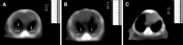
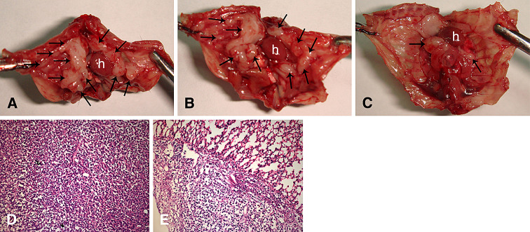
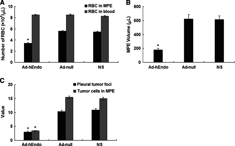
Methods: We developed a novel mouse model of MPE by injecting Lewis lung carcinoma (LLC) cells directly into pleural cavity of C57BL/6 mice. To evaluate the therapeutic effects of endostatin in this MPE model, we injected the Ad-hEndo into the pleural cavity of MPE-bearing mice three times with the 3-day interval.
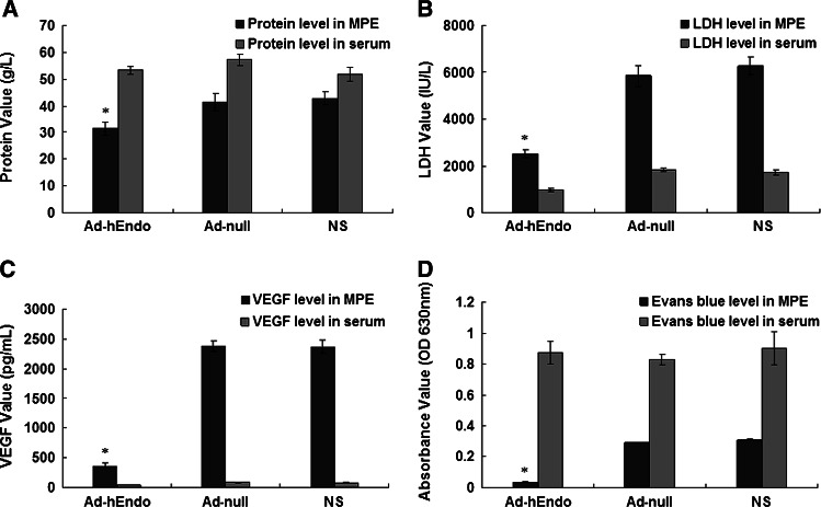
Results: We found that this treatment resulted in significant reduction in pleural effusion volume, the number of pleural tumor foci, microvessel density, and vascular permeability, while it significantly prolonged the survival time. In addition, VEGF level of MPE in the group administered with the Ad-hEndo was obviously decreased as compared with that in the two control groups administered with null-adenovirus (Ad-null) or normal saline.
Conclusions: Our work provides a rationale for future studies toward evaluating the effectiveness of the adenovirus-based endostatin therapy for MPE.
Figures


Similar articles
-
Recombinant human endostatin endostar suppresses angiogenesis and lymphangiogenesis of malignant pleural effusion in mice.PLoS One. 2012;7(12):e53449. doi: 10.1371/journal.pone.0053449. Epub 2012 Dec 28. PLoS One. 2012. PMID: 23285296 Free PMC article.
-
Effect of intrapleural anti-Vascular Endothelial Growth Factor (VEGF) associated with nab paclitaxel in a murine model of malignant pleural effusion.BMC Cancer. 2025 Aug 2;25(1):1261. doi: 10.1186/s12885-025-14622-x. BMC Cancer. 2025. PMID: 40753384 Free PMC article.
-
Development and Characterization of a Murine Lung Adenocarcinoma Cell Line With High Thoracic Pleural Metastatic Potential.In Vivo. 2025 Sep-Oct;39(5):2669-2680. doi: 10.21873/invivo.14067. In Vivo. 2025. PMID: 40877194 Free PMC article.
-
Interventions for the management of malignant pleural effusions: a network meta-analysis.Cochrane Database Syst Rev. 2016 May 8;2016(5):CD010529. doi: 10.1002/14651858.CD010529.pub2. Cochrane Database Syst Rev. 2016. Update in: Cochrane Database Syst Rev. 2020 Apr 21;4:CD010529. doi: 10.1002/14651858.CD010529.pub3. PMID: 27155783 Free PMC article. Updated.
-
Anti-vascular endothelial growth factor for diabetic macular oedema: a network meta-analysis.Cochrane Database Syst Rev. 2017 Jun 22;6(6):CD007419. doi: 10.1002/14651858.CD007419.pub5. Cochrane Database Syst Rev. 2017. Update in: Cochrane Database Syst Rev. 2018 Oct 16;10:CD007419. doi: 10.1002/14651858.CD007419.pub6. PMID: 28639415 Free PMC article. Updated.
Cited by
-
Effects of Combined Simultaneous and Sequential Endostar and Cisplatin Treatment in a Mice Model of Gastric Cancer Peritoneal Metastases.Gastroenterol Res Pract. 2017;2017:2920384. doi: 10.1155/2017/2920384. Epub 2017 Jan 19. Gastroenterol Res Pract. 2017. PMID: 28197204 Free PMC article.
-
Malignant pleural effusion: tumor-host interactions unleashed.Am J Respir Crit Care Med. 2012 Sep 15;186(6):487-92. doi: 10.1164/rccm.201203-0465PP. Epub 2012 May 31. Am J Respir Crit Care Med. 2012. PMID: 22652027 Free PMC article. Review.
-
Effect of Endostar combined with angiopoietin-2 inhibitor on malignant pleural effusion in mice.Med Oncol. 2015 Jan;32(1):410. doi: 10.1007/s12032-014-0410-0. Epub 2014 Dec 6. Med Oncol. 2015. PMID: 25479942
-
[Therapeutical effects of pleural injecting recombinant human endostain to malignant pleural effusion nude mice model].Zhongguo Fei Ai Za Zhi. 2015 May;18(5):266-71. doi: 10.3779/j.issn.1009-3419.2015.05.03. Zhongguo Fei Ai Za Zhi. 2015. PMID: 25975296 Free PMC article. Chinese.
-
Recombinant human endostatin endostar suppresses angiogenesis and lymphangiogenesis of malignant pleural effusion in mice.PLoS One. 2012;7(12):e53449. doi: 10.1371/journal.pone.0053449. Epub 2012 Dec 28. PLoS One. 2012. PMID: 23285296 Free PMC article.
References
-
- Antony VB, Loddenkemper R, Astoul P, Boutin C, Goldstraw P, Hott J et al (2000) Management of malignant pleural effusions. Am J Respir Crit Care Med 162:1987–2001 - PubMed
-
- Burrows CM, Mathews WC, Colt HG (2000) Predicting survival in patients with recurrent symptomatic malignant pleural effusions. Chest 117:73–78. doi:10.1378/chest.117.1.73 - PubMed
-
- Carmeliet P, Ferreira V, Breier G, Pollefeyt S, Kieckens L, Gertsenstein M et al (1996) Abnormal blood vessel development and lethality in embryos lacking a single VEGF allele. Nature 380:435–439. doi:10.1038/380435a0 - PubMed
-
- Ceyhan BB, Ozgun S, Celikel T, Yalcin M, Koc M (1996) IL-8 in pleural effusion. Respir Med 90:215–221. doi:10.1016/S0954-6111(96)90290-5 - PubMed
-
- Cheng D, Lee YCG, Rogers JT, Perkett EA, Moyers JP, Rodriguez RM et al (2000) Vascular endothelial growth factor level correlates with transforming growth factor-β isoform levels in pleural effusions. Chest 118:1747–1753. doi:10.1378/chest.118.6.1747 - PubMed
Publication types
MeSH terms
Substances
LinkOut - more resources
Full Text Sources
