

Regulation of the extracellular antioxidant selenoprotein plasma glutathione peroxidase (GPx-3) in mammalian cells
- PMID: 19219623
- PMCID: PMC2693281
- DOI: 10.1007/s11010-009-0049-x
Regulation of the extracellular antioxidant selenoprotein plasma glutathione peroxidase (GPx-3) in mammalian cells
Abstract
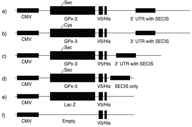
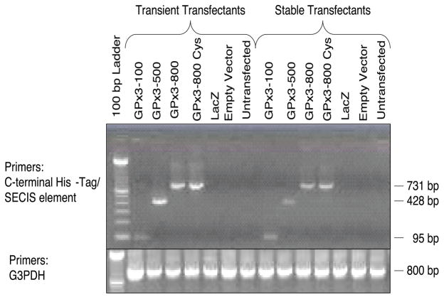
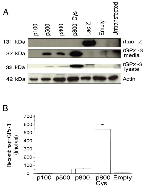
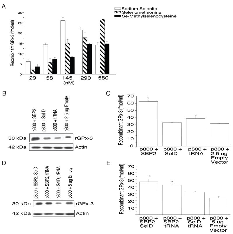
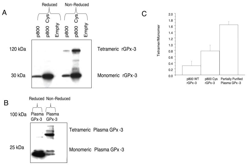
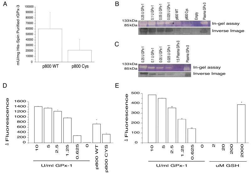
Plasma glutathione peroxidase (GPx-3) is a selenocysteine-containing extracellular antioxidant protein that catalyzes the reduction of hydrogen peroxide and lipid hydroperoxides. Selenoprotein expression involves the alternate recognition of a UGA codon as a selenocysteine codon and requires signals in the 3'-untranslated region (UTR), including a selenocysteine insertion sequence (SECIS), as well as specific translational cofactors. To ascertain regulatory determinants of GPx-3 expression and function, we generated recombinant GPx-3 (rGPX-3) constructs with various 3'-UTR, as well as a Sec73Cys mutant. In transfected Cos7 cells, the Sec73Cys mutant was expressed at higher levels than the wild type rGPx-3, although the wild type rGPx-3 had higher specific activity, similar to plasma purified GPx-3. A 3'-UTR with only the SECIS was insufficient for wild type rGPx-3 protein expression. Selenocompound supplementation and co-transfection with SECIS binding protein 2 increased wild type rGPx-3 expression. These results demonstrate the importance of translational mechanisms in GPx-3 expression.
Figures
Similar articles
-
Aminoglycosides decrease glutathione peroxidase-1 activity by interfering with selenocysteine incorporation.J Biol Chem. 2006 Feb 10;281(6):3382-8. doi: 10.1074/jbc.M511295200. Epub 2005 Dec 14. J Biol Chem. 2006. PMID: 16354666 Free PMC article.
-
Characterization of the UGA-recoding and SECIS-binding activities of SECIS-binding protein 2.RNA Biol. 2014;11(11):1402-13. doi: 10.1080/15476286.2014.996472. RNA Biol. 2014. PMID: 25692238 Free PMC article.
-
SBP, SECIS binding protein, binds to the RNA fragment upstream of the Sec UGA codon in glutathione peroxidase mRNA.Mol Biol Rep. 2000 Jun;27(2):99-105. doi: 10.1023/a:1007136123902. Mol Biol Rep. 2000. PMID: 11092556
-
The subcellular location of selenoproteins and the impact on their function.Nutrients. 2015 May 22;7(5):3938-48. doi: 10.3390/nu7053938. Nutrients. 2015. PMID: 26007340 Free PMC article. Review.
-
Chemical Biology Approaches to Interrogate the Selenoproteome.Acc Chem Res. 2019 Oct 15;52(10):2832-2840. doi: 10.1021/acs.accounts.9b00379. Epub 2019 Sep 16. Acc Chem Res. 2019. PMID: 31523956 Free PMC article. Review.
Cited by
-
Nutraceuticals: A New Challenge against Cadmium-Induced Testicular Injury.Nutrients. 2022 Feb 4;14(3):663. doi: 10.3390/nu14030663. Nutrients. 2022. PMID: 35277022 Free PMC article. Review.
-
The human selenoproteome: recent insights into functions and regulation.Cell Mol Life Sci. 2009 Aug;66(15):2457-78. doi: 10.1007/s00018-009-0032-4. Epub 2009 Apr 28. Cell Mol Life Sci. 2009. PMID: 19399585 Free PMC article. Review.
-
Catalytic Antioxidants in the Kidney.Antioxidants (Basel). 2021 Jan 18;10(1):130. doi: 10.3390/antiox10010130. Antioxidants (Basel). 2021. PMID: 33477607 Free PMC article. Review.
-
GPX3-Mediated Oxidative Stress Affects Pyrimidine Metabolism Levels in Stomach Adenocarcinoma via the AMPK/mTOR Pathway.Int J Clin Pract. 2024 Jan 30;2024:6875417. doi: 10.1155/2024/6875417. eCollection 2024. Int J Clin Pract. 2024. PMID: 38322113 Free PMC article.
-
Extracellular Glutathione Peroxidase GPx3 and Its Role in Cancer.Cancers (Basel). 2020 Aug 6;12(8):2197. doi: 10.3390/cancers12082197. Cancers (Basel). 2020. PMID: 32781581 Free PMC article. Review.
References
-
-
www.expasy.org .
-
-
- Behne D, Kyriakopoulos A. Mammalian selenium-containing proteins. Annu Rev Nutr. 2001;21:453–473. - PubMed
-
- Ghyselinck NB, Dufaure I, Lareyre JJ, Rigaudiere N, Mattei MG, Dufaure JP. Structural organization and regulation of the gene for the androgen-dependent glutathione peroxidase-like protein specific to the mouse epididymis. Mol Endocrinol. 1993;7:258–272. - PubMed
-
- Yoshimura S, Suemizu H, Taniguchi Y, Arimori K, Kawabe N, Moriuchi T. The human plasma glutathione peroxidase-encoding gene: organization, sequence and localization to chromosome 5q32. Gene. 1994;145:293–297. - PubMed
Publication types
MeSH terms
Substances
Grants and funding
LinkOut - more resources
Full Text Sources
Other Literature Sources
Molecular Biology Databases
Miscellaneous
