Review
. 2009 Jul-Aug;12(4):253-74.
doi: 10.2350/09-01-0586.1.

Genetic disorders of surfactant dysfunction

Affiliations
Review

Genetic disorders of surfactant dysfunction

Susan E Wert et al. Pediatr Dev Pathol. 2009 Jul-Aug.

Abstract

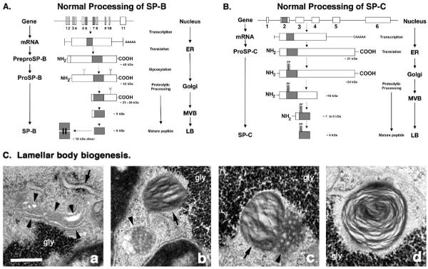
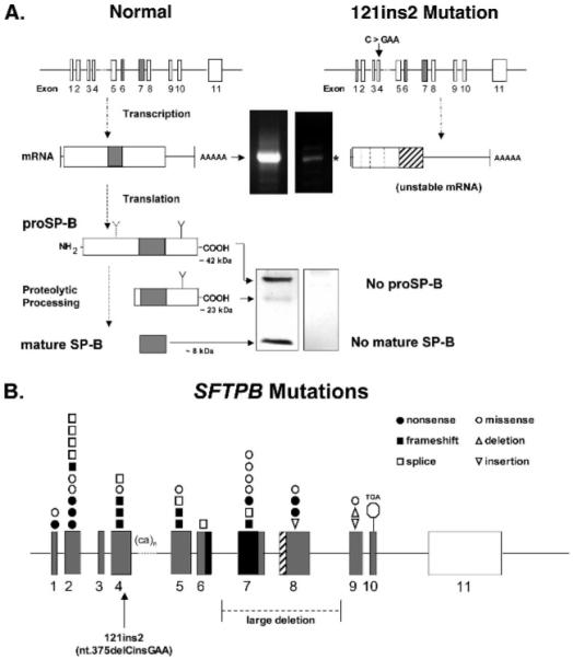
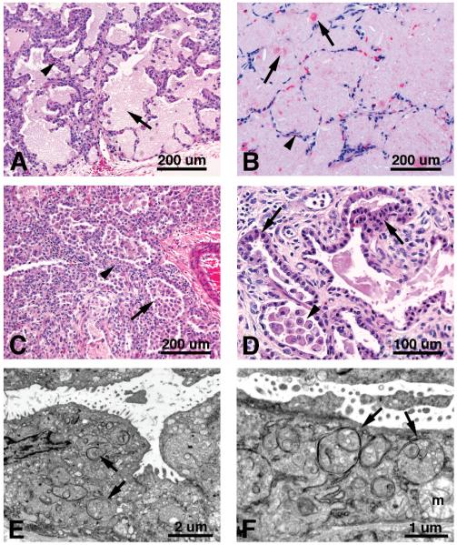
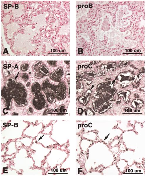
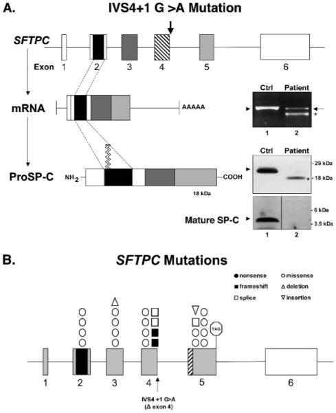
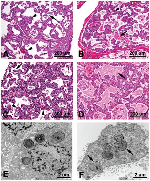
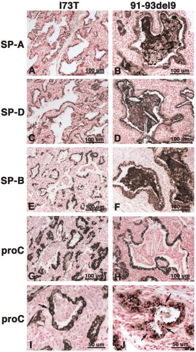
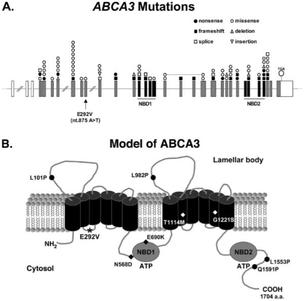
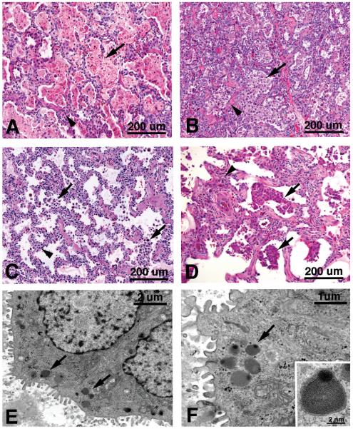
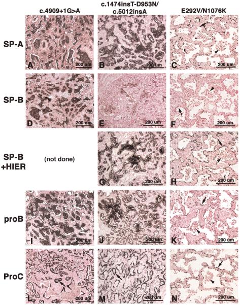
Mutations in the genes encoding the surfactant proteins B and C (SP-B and SP-C) and the phospholipid transporter, ABCA3, are associated with respiratory distress and interstitial lung disease in the pediatric population. Expression of these proteins is regulated developmentally, increasing with gestational age, and is critical for pulmonary surfactant function at birth. Pulmonary surfactant is a unique mixture of lipids and proteins that reduces surface tension at the air-liquid interface, preventing collapse of the lung at the end of expiration. SP-B and ABCA3 are required for the normal organization and packaging of surfactant phospholipids into specialized secretory organelles, known as lamellar bodies, while both SP-B and SP-C are important for adsorption of secreted surfactant phospholipids to the alveolar surface. In general, mutations in the SP-B gene SFTPB are associated with fatal respiratory distress in the neonatal period, and mutations in the SP-C gene SFTPC are more commonly associated with interstitial lung disease in older infants, children, and adults. Mutations in the ABCA3 gene are associated with both phenotypes. Despite this general classification, there is considerable overlap in the clinical and histologic characteristics of these genetic disorders. In this review, similarities and differences in the presentation of these disorders with an emphasis on their histochemical and ultrastructural features will be described, along with a brief discussion of surfactant metabolism. Mechanisms involved in the pathogenesis of lung disease caused by mutations in these genes will also be discussed.


Figures

Figure 1
Figure 1
Proposed structure, posttranslational processing, and trafficking of surfactant proteins B and C (SP-B and SP-C) in alveolar type II cells. A. Normal processing of SP-B. Human SP-B is encoded by a single gene, SFTPB, on chromosome 2, spanning about 10 kilobases (kb), and contains 10 introns (straight lines) and 11 exons (boxes), of which the last is untranslated. The mature SP-B peptide is encoded in exons 6 and 7 (shaded boxes). SFTPB is transcribed into a 2-kb mRNA that is translated in the endoplasmic reticulum (ER) to yield a 40-kd, 381-amino acid preproprotein. After cotranslational cleavage of its signal peptide, proSP-B undergoes glycosylation to yield a 42-kd intermediate form of the proprotein. Proteolytic cleavage of the NH2 and COOH termini of proSP-B occurs during trafficking of the proprotein through the Golgi apparatus and the multivesicular body (MVB), generating 23- to 26-kd and then 9-kd intermediate forms of the proprotein, before yielding the 8-kd, 79-amino acid, mature peptide that forms homodimers of ~18 kd in the mature lamellar body (LB). The shaded region of the proprotein represents the mature SP-B peptide. (Adapted from data in references 34–37, 43–45, and 48–53.) B. Normal processing of SP-C. Human SP-C is encoded by a single gene, SFTPC, on chromosome 8, spanning about 3.5 kb, and contains 5 introns (straight lines) and 6 exons (boxes), of which the last is untranslated. The mature SP-C peptide is encoded in exon 2 (shaded box). SFTPC is transcribed into a 0.9-kb messenger RNA that is translated in the ER to yield a 21-kd, 191- to 197-amino acid proprotein (proSP-C). ProSP-C then undergoes palmitoylation (PP) of cysteine residues within the mature peptide domain to yield a 24-kd intermediate. Proteolytic cleavage of the COOH and NH2 termini of proSP-C occurs during trafficking of the proprotein through the Golgi apparatus and the MVB, generating 16-kd and then 7- to 6-kd intermediate forms of the proprotein, before yielding the 4-kd, 35-amino acid, mature peptide that is found in the mature LB. The shaded region of the proprotein represents the mature SP-C peptide. (Adapted from data in references 38–42, 46, and 47.) C. Cellular components required for biosynthesis and processing of SP-B and SP-C. a. SP-B and SP-C are transcribed on ribosomes associated with the ER (arrows) and transported to the Golgi apparatus (arrowheads) for proteolytic processing of the larger precursor proteins to smaller, intermediate forms of the proproteins. b. Endosomal vesicles containing proSP-B and proSP-C bud off the Golgi apparatus and accumulate in MVBs (arrowheads). An LB with prominent phospholipid lamellae is seen nearby (arrow). c. The partially processed proproteins are delivered to LBs via fusion of the MVB (arrowhead) with the LB (arrow) to form a composite body, wherein final processing of the proproteins to mature SP-B and SP-C is thought to occur. d. The mature LB is composed of both surfactant phospholipids and the fully processed, mature SP-B and SP-C peptides that are now tightly associated with the phospholipid bilayers. Images were acquired by electron microscopy of newly forming LBs in immature alveolar type II cells found in fetal mouse lung tissue. gly, glycogen. Scale bar = 500 nm.
Figure 2
Figure 2
Structural models of the human surfactant protein B (SP-B) gene and protein illustrating abnormal processing of SP-B and the location of mutations found throughout the gene. A. Comparison of normal and abnormal transcription, synthesis, and processing of SP-B caused by the 121ins2 mutation. Aberrant transcription of SFTPB caused by the 121ins2 mutation results in formation of an unstable messenger RNA (mRNA) that is rapidly degraded and barely detected by polymerase chain reaction (PCR) analysis (faint white band on black at *, upper right panel) compared with the presence of normal mRNA observed in a control patient (white band on black at arrow, upper left panel). This results in the complete absence of both proprotein SP-B (proSP-B) and the mature SP-B peptide, as assessed by immunoblot of protein isolated from lung tissue of an affected patient (no black bands, lower right panel). In contrast, the presence of 42-kd and 23-kd forms of proSP-B, as well as the 8-kd mature SP-B peptide (black bands on white at arrows, lower left panel) can be detected in protein isolated from lung tissue of a control patient. B. Structural model of SFTPB illustrating the location of mutations found throughout the gene. The mature peptide is encoded in exons 6 and 7 (black boxes); exon 11 (white box) is not translated. Mutations in SFTPB include a variety of nonsense, missense, frameshift, and splice-site mutations, as well as insertions and deletions. The common mutation, 121ins2, is located in exon 4 (arrow). A large deletion, encompassing exons 7 and 8, has been reported recently. TGA, translational stop sequence located in exon 10.
Figure 3
Figure 3
Histopathology and ultrastructural features of disorders caused by mutations in the human surfactant protein B (SP-B) gene. A. Autopsy tissue from a 23-day-old child who was homozygous for the 121ins2 mutation. Alveolar proteinosis with foamy, eosinophilic, lipoproteinaceous material filling the alveoli (arrow) is typically found in the lung of patients with SP-B mutations. Thickened alveolar septa (arrowheads) are also a prominent feature of this disorder. B. Biopsy tissue from a 6-year-old child with a granulocyte-macrophage colony-stimulating factor receptor alpha chain mutation [129]. In contrast to SP-B deficiency, the large amount of alveolar proteinosis material found in this genetic disorder is denser and contains larger globules of eosinophilic material (arrows), which fills and expands the alveoli. There is also good preservation of the alveolar septa (arrowhead). C. Explant tissue from a 13-month-old child who was a compound heterozygote for the 121ins2 and C100G mutations [107], demonstrating infantile desquamative interstitial pneumonitis (DIP) with accumulation of foamy alveolar macrophages in the alveoli (arrow) and little to no alveolar proteinosis. The alveolar epithelia are hyperplastic, and the thickened alveolar septa (arrowhead) contain lymphocytic infiltrates in this sample. D. Autopsy tissue from a 2-month-old child who was a compound heterozygote for the 121ins2 and c.282-2delA mutations [107]. Higher magnification of hyperplastic alveolar epithelia with prominent type II cells (arrows) and accumulation of foamy macrophages in the alveolar lumen is shown. Note the prominent interstitial widening composed of loose connective tissue and disruption of the normal alveolar capillary architecture, which precludes normal gas exchange and lends an immature appearance to the lung. (Hematoxylin and eosin-stained paraffin sections.) E. In lieu of normal lamellar bodies, electron microscopic analysis demonstrates the presence of many large, membrane-bound structures containing smaller membranous vesicles (arrows) and, occasionally, several concentric layers of phospholipid lamellae in the type II cells of a child with the 121ins2 mutation. F. Higher magnification of these aberrant structures (arrows), which are similar in appearance to the multivesicular and composite bodies found during lamellar body biogenesis. m, disrupted mitochondria.
Figure 4
Figure 4
Immunohistochemical staining for the surfactant proteins in lung tissue from subjects with mutations in the human surfactant protein B (SP-B) gene. Autopsy tissue from a child who was homozygous for the 121ins2 mutation is shown in panels A through D; biopsy tissue from a control sample is shown in panels E and F. No immunostaining for mature SP-B (A) or proprotein SP-B (proSP-B; B) is found in the 121ins2 mutation. On the other hand, the alveolar proteinosis material (arrow) found in this mutation stains intensely for SP-A (C) and for proSP-C (D), which are detected in both alveolar type II cells (black reaction product at arrowhead) and in the alveolar lumen (black reaction product at arrow). In contrast, immunostaining for mature SP-B (E) and proSP-C (F) is restricted to alveolar type II cells (arrows) and is not detected in the alveolar lumen in biopsy tissue from a control lung. Immunohistochemistry was performed using polyclonal antibodies to (1) full-length SP-A, (2) the mature SP-B peptide, (3) the carboxyl terminus of proSP-B, and (4) the amino terminus of proSP-C. A color version of this figure is available online.
Figure 5
Figure 5
Structural models of the human surfactant protein C (SP-C) gene and protein illustrating abnormal processing of SP-C and the location of mutations found throughout the gene. A. Abnormal processing of SP-C caused by the IVS4+1G>A mutation. The mutation, IVS4+1G>A (also known as c.460+1G>A or Δexon 4), is located at the junction of exon 4 and its adjacent intron (arrow), and is found on only one allele of the affected patient. This causes exon 4 to be skipped, resulting in a truncated form of the messenger RNA for this allele, which contains sequences for exons 1 through 3 and exon 5. This can be observed by reverse transcriptase polymerase chain reaction analysis of the patient’s RNA (lower band at *, lane 2, upper panel). A normal-sized band (upper band at arrow, lane 2, upper panel) indicating that the nonmutated allele is found in the affected patient, as well as in a control (Ctrl) patient (upper band at arrowhead, lane 1, upper panel). This results in translation of an aberrant 18-kd form of the proprotein (lower band at *, lane 2, middle panel depicting an immunoblot of the protein) compared with the 21-kd proprotein observed in the control patient (upper band at arrowhead, lane 1, middle panel). No mature peptide is found by immunoblot in the affected patient (lane 2, right lower panel of protein blot) when compared with the control patient (band at arrowhead, lane 1, left lower panel). B. Structural model of SFTPC illustrating the location of mutations found throughout the gene. Mutations in SFTPC include a variety of nonsense, missense, frameshift, and splice-site mutations, as well as insertions and deletions. The IVS4+1G>A mutation (also known as c.460+1G>A or the Δexon 4 mutation) is located at the junction of exon 4 and its adjacent intron (arrow). The mature peptide is encoded in exon 2 (black boxes); exon 6 (white box) is not translated. TAG, translational stop sequence located in exon 10.
Figure 6
Figure 6
Histopathology and ultrastructural features of genetic disorders caused by mutations in the human surfactant protein C (SP-C) gene. A. Nonspecific interstitial pneumonitis (NSIP), with thickened alveolar septa (arrowhead) and a few alveolar macrophages, is seen in a biopsy from a 6-week–old child, heterozygous for the Δexon 4 mutation (also known as IVS4+1 G>A or c.460+1G>A) [143]. B. Chronic pneumonitis of infancy (CPI) with alveolar septal thickening (arrowheads) and accumulation of large, foamy macrophages and granular, eosinophilic, alveolar proteinosis material in the alveoli (arrow) is seen in a biopsy from a 9-month–old child, heterozygous for the P115L mutation [144]. C. Another example of CPI, with diffuse alveolar septal thickening, alveolar type II cell hyperplasia, and accumulation of macrophages (arrow) in the alveolar lumen, is seen in a biopsy from a 1-year–old child, heterozygous for the common I73T mutation [151]. Muscularization of the alveolar septa and ducts (arrowhead) and inflammatory cell infiltrates are found in the adjacent thickened interstitial structures. D. Accumulation of larger amounts of foamy alveolar proteinosis material with cholesterol clefts (arrow) is found in explanted tissue from a 9-month-old child, heterozygous for the 91-93del9 mutation [148]. (Hematoxylin and eosin-stained paraffin sections.) E. Electron microscopic analysis demonstrates the presence of large, well-organized lamellar bodies (arrows) found in the type II cells of a 6-week-old child heterozygous for the Δexon 4 mutation. F. Large composite bodies containing multiple lamellar body–like structures and membrane-bound vesicles were also found in this sample (arrows).
Figure 7
Figure 7
Immunohistochemical staining for the surfactant proteins in lung tissue from subjects with mutations in the human surfactant protein C (SP-C) gene. Immunohistochemistry for the common I73T mutation [151] is shown in column one (A, C, E, G, and I) and for the 91-93del9 mutation [148] in column two (B, D, F, H, and J). Immunostaining for SP-A (A, B), SP-D, (C, D), mature SP-B (E, F), and proprotein SP-C (proSP-C; G through J) is robust in both mutations (black reaction product). The alveolar proteinosis material and macrophages found in the 91-93del9 mutation are immunopositive for SP-A (B), SP-D (D), and mature SP-B (F), but not for proSP-C (H), which is restricted to the alveolar type II cells. Two different immunostaining patterns are detected for proSP-C. Diffuse staining of the alveolar type II cell cytoplasm is seen in the I73T mutation (I), while a more perinuclear staining pattern is seen in the 91-93del9 mutation (J). Immunohistochemistry was performed using polyclonal antibodies to (1) full-length SP-A, (2) full-length SP-D, (3) the mature SP-B peptide, (4) the carboxyl terminus of proSP-B, and (5) the amino terminus of proSP-C. A color version of this figure is available online.
Figure 8
Figure 8
Structural models of the human ABCA3 gene and protein illustrating the location of mutations found throughout the gene and protein. A. Structural model of the ABCA3 gene. ABCA3 is encoded by a single gene, ABCA3, on chromosome 16 (16p13.3), and contains 33 exons (boxes). The adenosine triphosphate (ATP)-binding domain or nucleotide-binding domain (NBD) is encoded in exons 14–17 (NBD1) and exons 27–30 (NBD2) (black boxes). Mutations in ABCA3 include a variety of nonsense, missense, frameshift, and splice-site mutations, as well as insertions and deletions. The common mutation, E292V, is located in exon 9 (arrow). TGA, translational stop sequence located in exon 33. B. Structural model of the ABCA3 protein. ABCA3 is a 1704-amino acid, integral membrane protein located at the limiting membrane of the lamellar body (LB). It contains 2 homologous repeats, each consisting of 6 putative transmembrane helices and an ATP-binding domain. ABCA3 is thought to be oriented in the lipid bilayer such that its ATP-binding domains (NBD1, NBD2) are located in the cytoplasm. Phospholipids are then actively transported from the cytosol to the interior of the LB through the membrane channel (black cylinders) that is formed in the phospholipid bilayer. The location of mutations that have been shown to impair ABCA3 function [191,192], either by interfering with ATP hydrolysis (N568D, E690K, T1114M, G1221S) or with trafficking of the protein from the endoplasmic reticulum (ER) to the LB (L101P, L982P, L1553P, Q1591P), are illustrated. *, location of the E292V mutation. (Adapted from Matsumura and colleagues [191,192].)
Figure 9
Figure 9
Histopathology and ultrastructural features of disorders caused by mutations in the human ABCA3 gene. A. Alveolar proteinosis, admixed with alveolar macrophages, and thickened alveolar septa (arrowhead) are seen in a biopsy from a neonate with fatal lung disease who was homozygous for the c.4909+1G>A splicing mutation [173]. B. Infantile desquamative interstitial pneumonitis (DIP) with accumulation of foamy macrophages in the alveoli is seen in an autopsy from 33-day-old child who was a compound heterozygote for the c.1474insT-D953N and c.5012insA mutations. The alveolar epithelium is hyperplastic, and muscularization of the alveolar septa is found in the adjacent thickened interstitial structures (arrowhead). C. Thickened alveolar septa (arrowhead) and accumulation of alveolar macrophages (arrow) are seen in explanted tissue from a 21-year-old adult with a diagnosis of DIP and who is a compound heterozygote for the E292V and N1076K mutations [176]. D. Lung remodeling with thickened alveolar septa and macrophage accumulation are seen in a biopsy from a 2-year-old child with prolonged survival (>12 years old) who is a compound heterozygote for the E292V and c.1742-9G>A mutations [176]. (Hematoxylin and eosin-stained paraffin sections.) E. Electron microscopic analysis demonstrates the presence of small, abnormal, lamellarlike bodies (arrows) with eccentrically placed electron-dense inclusions in a biopsy from a neonate who was homozygous for the c.4909+1G>A mutation (see panel A). F. Higher magnification of small, lamellarlike bodies (arrow) with tightly packed phospholipid membranes (inset) found in the type II cells of this sample.
Figure 10
Figure 10
Immunohistochemical staining for the surfactant proteins in lung tissue from subjects with mutations in the human ABCA3 gene. Immunohistochemistry from a neonate, homozygous for the c.4909+1G>A mutation [173], is shown in column one (A, D, I, and L); from a 33-day-old, compound heterozygote for the c.1474insT-D953N and c.5012insA mutations, is shown in column two (B, E, G, J, and M); and from a 21-year-old adult, compound heterozygote for the E292V and N1076K mutations [176], is shown in column 3 (C, F, H, K, and N). Immunostaining for surfactant protein A (SP-A; A, B, and C), proprotein SP-B (proSP-B; I, J, and K), and proSP-C (L, M, and N) is robust in all three mutations (black reaction product). Immunostaining for mature SP-B is readily detected in the c.4909+1G>A mutation (D), but is weak or not detected in the other two mutations (E, F). Immunostaining for mature surfactant protein B (SP-B) is recovered after use of heat-induced epitope retrieval (G, H). The alveolar proteinosis material and macrophages found in the first 2 mutations are immunopositive for SP-A (A, B), SP-B (D, G), and proSP-B (I, J). Immunostaining for proSP-C, however, is restricted to alveolar type II cells (arrows) in all three mutations (L, M, and N). Immunostaining for the surfactant proteins is detected primarily in the alveolar type II cells (arrows) in the E292V/N1076K mutation (C, F, H, K, and N). There is little to no secreted, immunopositive, proteinosis material found in this sample, and no immunostaining is detected in the alveolar macrophages (arrowheads). Immunohistochemistry was performed using polyclonal antibodies to (1) full-length SP-A, (2) the mature SP-B peptide, (3) the carboxyl terminus of proSP-B, and (4) the amino terminus of proSP-C. A color version of this figure is available online.

Similar articles

Cited by

References

    1. Nogee LM. Genetics of the hydrophobic surfactant proteins. Biochim Biophys Acta. 1998;1408:323–333. - PubMed
    1. Whitsett JA, Weaver TE. Hydrophobic surfactant proteins in lung function and disease. N Engl J Med. 2002;347:2141–2148. - PubMed
    1. Nogee LM. Genetic mechanisms of surfactant deficiency. Biol Neonate. 2004;85:314–318. - PubMed
    1. Nogee LM. Alterations in SP-B and SP-C expression in neonatal lung disease. Annu Rev Physiol. 2004;66:601–623. - PubMed
    1. Hartl D, Griese M. Interstitial lung disease in children–genetic background and associated phenotypes. Respir Res. 2005;6:32–48. - PMC - PubMed

MeSH terms

Substances