

Theory of sampling and its application in tissue based diagnosis
- PMID: 19220904
- PMCID: PMC2649041
- DOI: 10.1186/1746-1596-4-6
Theory of sampling and its application in tissue based diagnosis
Abstract
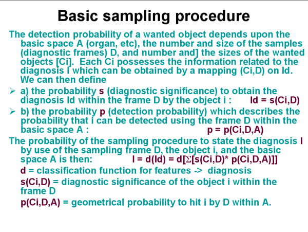
Background: A general theory of sampling and its application in tissue based diagnosis is presented. Sampling is defined as extraction of information from certain limited spaces and its transformation into a statement or measure that is valid for the entire (reference) space. The procedure should be reproducible in time and space, i.e. give the same results when applied under similar circumstances. Sampling includes two different aspects, the procedure of sample selection and the efficiency of its performance. The practical performance of sample selection focuses on search for localization of specific compartments within the basic space, and search for presence of specific compartments.
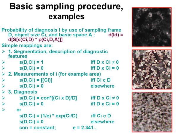
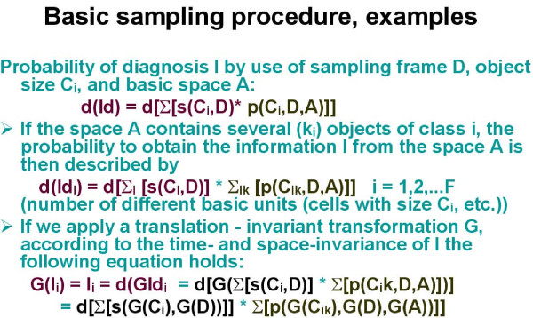
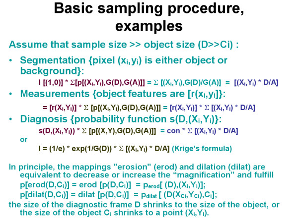
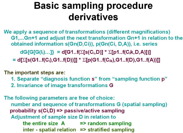
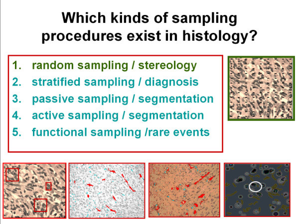
Methods: When a sampling procedure is applied in diagnostic processes two different procedures can be distinguished: I) the evaluation of a diagnostic significance of a certain object, which is the probability that the object can be grouped into a certain diagnosis, and II) the probability to detect these basic units. Sampling can be performed without or with external knowledge, such as size of searched objects, neighbourhood conditions, spatial distribution of objects, etc. If the sample size is much larger than the object size, the application of a translation invariant transformation results in Kriege's formula, which is widely used in search for ores. Usually, sampling is performed in a series of area (space) selections of identical size. The size can be defined in relation to the reference space or according to interspatial relationship. The first method is called random sampling, the second stratified sampling.
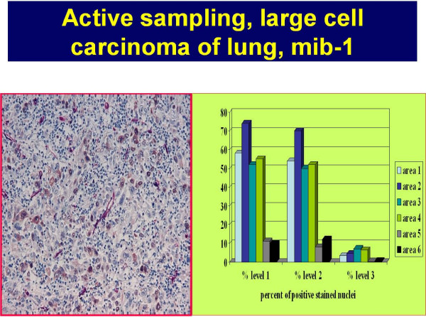
Results: Random sampling does not require knowledge about the reference space, and is used to estimate the number and size of objects. Estimated features include area (volume) fraction, numerical, boundary and surface densities. Stratified sampling requires the knowledge of objects (and their features) and evaluates spatial features in relation to the detected objects (for example grey value distribution around an object). It serves also for the definition of parameters of the probability function in so-called active segmentation.
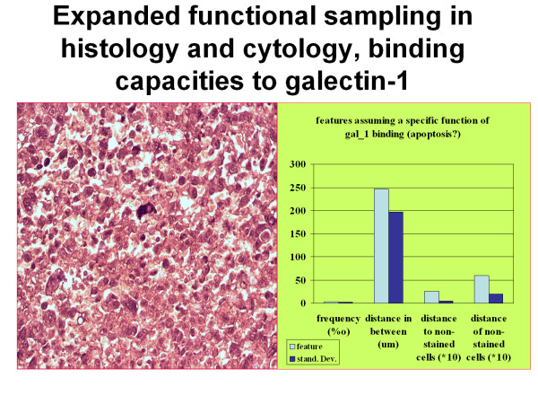
Conclusion: The method is useful in standardization of images derived from immunohistochemically stained slides, and implemented in the EAMUS system http://www.diagnomX.de. It can also be applied for the search of "objects possessing an amplification function", i.e. a rare event with "steering function". A formula to calculate the efficiency and potential error rate of the described sampling procedures is given.
Figures
Similar articles
-
How to measure image quality in tissue-based diagnosis (diagnostic surgical pathology).Diagn Pathol. 2008 Jul 15;3 Suppl 1(Suppl 1):S11. doi: 10.1186/1746-1596-3-S1-S11. Diagn Pathol. 2008. PMID: 18673499 Free PMC article.
-
Digitized pathology: theory and experiences in automated tissue-based virtual diagnosis.Rom J Morphol Embryol. 2006;47(1):21-8. Rom J Morphol Embryol. 2006. PMID: 16838053
-
AI (artificial intelligence) in histopathology--from image analysis to automated diagnosis.Folia Histochem Cytobiol. 2009 Jan;47(3):355-61. doi: 10.2478/v10042-009-0087-y. Folia Histochem Cytobiol. 2009. PMID: 20164018 Review.
-
Early object relations into new objects.Psychoanal Study Child. 2001;56:39-67; discussion 68-75. doi: 10.1080/00797308.2001.11800664. Psychoanal Study Child. 2001. PMID: 12102023
-
Methods in quantitative image analysis.Histochem Cell Biol. 1996 May;105(5):333-55. doi: 10.1007/BF01463655. Histochem Cell Biol. 1996. PMID: 8781988 Review.
Cited by
-
Optical coherence tomography holds promise to transform the diagnostic anatomic pathology gross evaluation process.J Biomed Opt. 2022 Sep;27(9):096003. doi: 10.1117/1.JBO.27.9.096003. J Biomed Opt. 2022. PMID: 36050827 Free PMC article.
-
Clinical validation of the gastrointestinal NET grading system: Ki67 index criteria of the WHO 2010 classification is appropriate to predict metastasis or recurrence.Diagn Pathol. 2013 Apr 22;8:65. doi: 10.1186/1746-1596-8-65. Diagn Pathol. 2013. PMID: 23607525 Free PMC article.
-
Unbiased analysis of obesity related, fat depot specific changes of adipocyte volumes and numbers using light sheet fluorescence microscopy.PLoS One. 2021 Mar 16;16(3):e0248594. doi: 10.1371/journal.pone.0248594. eCollection 2021. PLoS One. 2021. PMID: 33725017 Free PMC article.
-
Automatic measurement of epithelium differentiation and classification of cervical intraneoplasia by computerized image analysis.Diagn Pathol. 2010 Jan 22;5:7. doi: 10.1186/1746-1596-5-7. Diagn Pathol. 2010. PMID: 20148100 Free PMC article.
-
An experimental study of pathologist's navigation patterns in virtual microscopy.Diagn Pathol. 2010 Nov 18;5:71. doi: 10.1186/1746-1596-5-71. Diagn Pathol. 2010. PMID: 21087502 Free PMC article.
References
-
- Kayser G, Radziszowski D, Bzdyl P, et al. Theory and implementation of an electronic, automated measurement system for images obtained from immunohistochemically stained slides. Anal Quant Cytol Histol. 2006;28:27–38. - PubMed
-
- Kayser K, Molnar B, Weinstein R. Virtual Microscopy: Fundamentals, Applications, Perspectives of Electronic Tissue-based Diagnosis. Berlin: VSV Interdisciplinary Medical Publishing; 2006.
-
- Kayser K, Kayser G. Basic aspects of and recent developments in telepathology in Europe, with specific emphasis on quality assurance. Anal Quant Cytol Histol. 1999;21:319–328. - PubMed
LinkOut - more resources
Full Text Sources
Other Literature Sources
