

Palifermin-associated papular eruption
- PMID: 19221263
- PMCID: PMC2684027
- DOI: 10.1001/archdermatol.2008.548
Palifermin-associated papular eruption
Abstract
Background: Palifermin is a recombinant human keratinocyte growth factor that is used to reduce the duration and severity of oral mucositis in patients undergoing hematopoietic stem cell transplantation after myelotoxic therapy. Cutaneous adverse reactions associated with keratinocyte growth factor are reported to be rash, pruritus, and erythema.
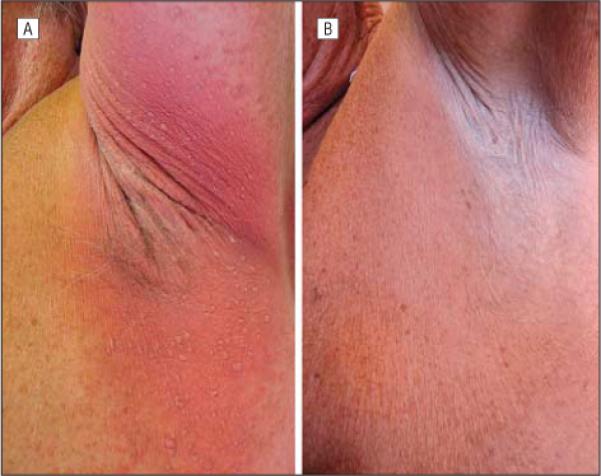
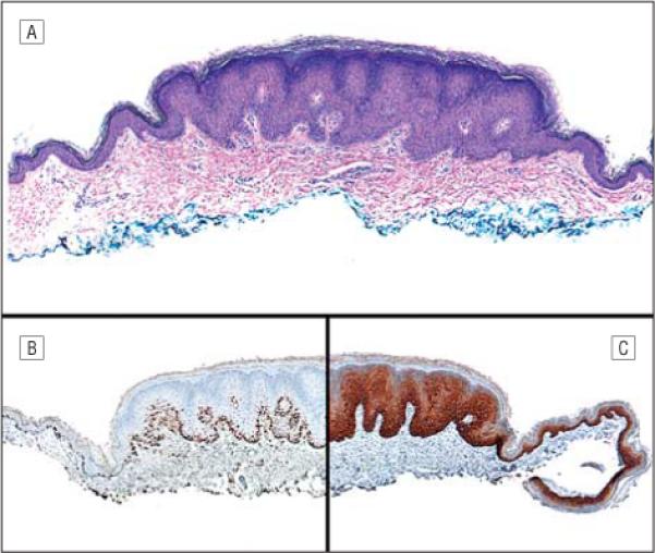
Observations: After receiving palifermin following autologous hematopoietic stem cell transplantation and treatment with melphalan, a patient developed erythema and lichenoid papules that were distributed primarily in intertriginous areas. A biopsy specimen of the papules showed a striking resemblance to verrucae, but in situ hybridization studies were negative for human papillomavirus. Immunohistochemical staining with antibodies to Ki-67 and cytokeratin 5/6 showed increased keratinocyte proliferation in lesional skin.
Conclusions: After treatment with palifermin, a papular eruption clinically resembling lichen planus or plane warts, with histologic features of verruca plana, and intertriginous erythema may occur. In this case, neither eruption required treatment, and spontaneous resolution was observed over days to weeks. Histopathologic staining patterns of Ki-67 and cytokeratin 5/6 may be useful in identifying adverse reactions to palifermin therapy.
Figures
References
-
- Farrell CL, Bready JV, Rex KL, et al. Keratinocyte growth factor protects mice from chemotherapy and radiation-induced gastrointestinal injury and mortality. Cancer Res. 1998;58(5):933–939. - PubMed
-
- Werner S. Keratinocyte growth factor: a unique player in epithelial repair processes. Cytokine Growth Factor Rev. 1998;9(2):153–165. - PubMed
-
- Zia-Amirhosseini P, Salfi M, Leese P, et al. Pharmacokinetics, pharmacodynamics, and safety assessment of palifermin (rHuKGF) in healthy volunteers. Clin Pharmacol Ther. 2006;79(6):558–569. - PubMed
-
- Spielberger R, Stiff P, Bensinger W, et al. Palifermin for oral mucositis after intensive therapy for hematologic cancers. N Engl J Med. 2004;351(25):2590–2598. - PubMed
Publication types
MeSH terms
Substances
Grants and funding
LinkOut - more resources
Full Text Sources
Medical
Research Materials
