. 2009 Mar 16;206(3):497-505.
doi: 10.1084/jem.20082129. Epub 2009 Feb 16.

Identification of antigen-presenting dendritic cells in mouse aorta and cardiac valves

Affiliations

Identification of antigen-presenting dendritic cells in mouse aorta and cardiac valves

Jae-Hoon Choi et al. J Exp Med. .

Abstract

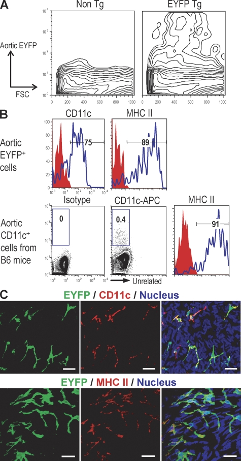
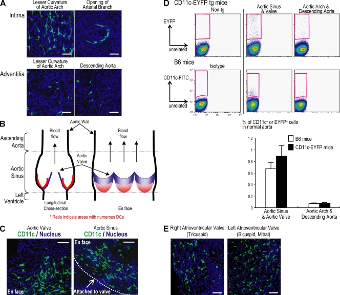
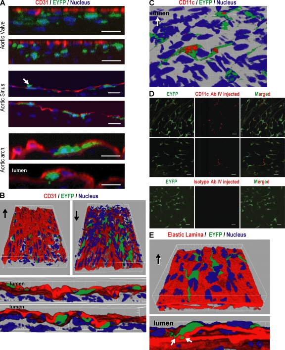
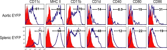
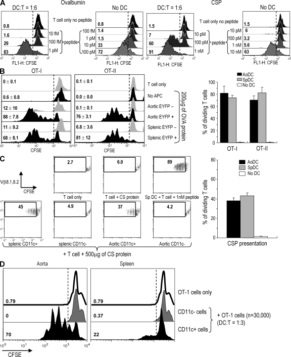
Presumptive dendritic cells (DCs) bearing the CD11c integrin and other markers have previously been identified in normal mouse and human aorta. We used CD11c promoter-enhanced yellow fluorescent protein (EYFP) transgenic mice to visualize aortic DCs and study their antigen-presenting capacity. Stellate EYFP(+) cells were readily identified in the aorta and could be double labeled with antibodies to CD11c and antigen-presenting major histocompatability complex (MHC) II products. The DCs proved to be particularly abundant in the cardiac valves and aortic sinus. In all aortic locations, the CD11c(+) cells localized to the subintimal space with occasional processes probing the vascular lumen. Aortic DCs expressed little CD40 but expressed low levels of CD1d, CD80, and CD86. In studies of antigen presentation, DCs selected on the basis of EYFP expression or binding of anti-CD11c antibody were as effective as DCs similarly selected from the spleen. In particular, the aortic DCs could cross-present two different protein antigens on MHC class I to CD8(+) TCR transgenic T cells. In addition, after intravenous injection, aortic DCs could capture anti-CD11c antibody and cross-present ovalbumin to T cells. These results indicate that bona fide DCs are a constituent of the normal aorta and cardiac valves.


Figures

Figure 1.
Figure 1.
Visualizing aortic DCs using CD11c-EYFP transgenic mice. (A) The presence of EYFP+ cells in aortic cell suspension from EYFP transgenic mice. Aortic segments from EYFP transgenic mice (n = 5) were pooled and dissociated by incubation with an enzyme mixture (see Materials and methods) and then subjected to flow cytometric analysis. (B) Experiments were performed as in A, but the aortic CD11c+ cells were from nontransgenic mice (n = 3) and compared for expression of CD11c and MHC II to aortic EYFP+ cells. (C) Immunohistochemical staining of CD11c and MHC II in aortic sheets. Each experiment was performed at least three times. Bars, 20 µm.
Figure 2.
Figure 2.
Predominant localization of aortic DCs in the aortic valve and sinus. Aortic segments from aortic sinus to diaphragm were opened longitudinally to make aortic sheets. The distribution of aortic DCs was analyzed using confocal microscopy. (A) EYFP+ DCs in the intima of the aortic arch (top left) and openings of arterial branches (top right). Adventitial EYFP+ DCs were scattered throughout the aorta including aortic arch (bottom left) and descending aorta (bottom right) (B) Diagram of aortic sinus. Red indicates DC-abundant areas. (C) EYFP+ DCs in aortic valve (left) and sinus (right). (D) Flow cytometric analysis of EYFP+ cells. The mean and SD of three experiments are shown below, with the aortic cell suspension in each experiment being from at least three mice. (E) The presence of DCs in normal cardiac valves including left (left panel) and right (right panel) atrioventricular valves. The cardiac valves were isolated using microscissors under guidance of a dissecting microscope. The valvular sheets were mounted on glass slides and observed by confocal microscopy. Each figure is representative of at least three experiments. Bars, 40 µm.
Figure 3.
Figure 3.
The anatomical position of aortic DCs in aortic wall. After immunostaining the aortic sheet using CD31 antibody to visualize the endothelium, confocal images were taken along the Z axis to visualize all dendritic processes from each EYFP+ DC and were reconstituted to a Z stack. (A) Reconstituted Z planes from aortic valve (top), aortic sinus (middle), and aortic arch (bottom) show that the aortic DCs are localized beneath the endothelium. A small number of DCs in the aortic sinus extend their processes into the vessel lumen (A, middle, arrow). (B) Three-dimensional images were reconstituted using the Imaris program to better visualize the position of aortic DCs. Arrows indicate the vessel lumen side. (C) Exposure of some dendritic processes into the lumen. The aortic sheets from EYFP transgenic mice were stained with CD11c antibody without tissue permeabilization to see whether dendritic processes could capture antibody from the lumen. The confocal images were reconstituted in three dimensions. (D) Biotinylated anti-CD11c and hamster IgG control antibody were injected i.v. to CD11c-EYFP mice. After 3 h, the aortic segments were isolated, incubated with horseradish peroxidase–conjugated streptavidin, and the stained CD11c was enhanced with Alexa Fluor 555 tyramide. (E) The elastic lamina was visualized using its autofluorescence. The confocal images were reconstituted in three dimensions. Each figure is representative of at least three experiments. Bars, 20 µm.
Figure 4.
Figure 4.
Comparison of cell surface markers of aortic and splenic DCs. Aortic and spleen cell suspensions from transgenic mice (n = 5) was stained with antibodies to each surface marker as shown. Each figure is representative of at least three experiments.
Figure 5.
Figure 5.
Antigen presentation to T cells by aortic DCs. (A) Comparison between OT-1 and YA26 TCR transgenic CD8+ T cells. The indicated concentrations of OT-1 peptide (SIINFEKL) or CS peptide (SYVPSAEQI) were added to each T cell with or without splenic DCs. After 4 d, T cell proliferation was analyzed by CFSE dilution. (B) Presentation to OVA-specific TCR transgenic T cells. Aortic and splenic EYFP+ and EYFP cells were isolated from EYFP transgenic mice (n = 10) by FACS sorting. CD8+ OT-I and CD4+ OT-II cells were isolated using Dynabeads. 200 µg/ml OVA protein was added to the DC–T cell cocultures. Proliferation of OT-I and OT-II cells was evaluated by CFSE dilution on the FACS. The right shows the mean and SD of three experiments. (C) Presentation to YA26 T cells. Aortic and splenic CD11chigh CD3 CD19 DX5 cells were isolated using FACS sorting and YA26 T cells with Dynabeads. 500 µg/ml CSP was added to the DC–T cell cocultures. Proliferation of CSP-specific TCR transgenic T cells was evaluated by CFSE dilution. The right shows the mean and SD of three experiments. (D) 5 mg OVA was injected i.v. to CD11c-EYFP (n = 10) mice. After 20 h, the aortic cell suspensions were prepared and sorted to isolate EYFP+ and EYFP cells. Spleen EYFP+ and EYFP cells were also isolated from the mice. The cells were added to OT-1 T cells at ratio of 1:3. After 3 d, T cell proliferation was analyzed by CFSE dilution. This result is representative of two independent experiments.

References

    1. Johnston S.L., Lock R.J., Gompels M.M. 2002. Takayasu arteritis: a review.J. Clin. Pathol. 55:481–486 - PMC - PubMed
    1. Ma-Krupa W., Kwan M., Goronzy J.J., Weyand C.M. 2005. Toll-like receptors in giant cell arteritis.Clin. Immunol. 115:38–46 - PubMed
    1. Shimizu K., Mitchell R.N., Libby P. 2006. Inflammation and cellular immune responses in abdominal aortic aneurysms.Arterioscler. Thromb. Vasc. Biol. 26:987–994 - PubMed
    1. Hansson G.K., Libby P. 2006. The immune response in atherosclerosis: a double-edged sword.Nat. Rev. Immunol. 6:508–519 - PubMed
    1. Steinman R.M., Hemmi H. 2006. Dendritic cells: translating innate to adaptive immunity.Curr. Top. Microbiol. Immunol. 311:17–58 - PubMed

Publication types

MeSH terms