. 2009 Mar 16;206(3):507-14.
doi: 10.1084/jem.20082113. Epub 2009 Feb 16.

A novel myelin P0-specific T cell receptor transgenic mouse develops a fulminant autoimmune peripheral neuropathy

Affiliations

A novel myelin P0-specific T cell receptor transgenic mouse develops a fulminant autoimmune peripheral neuropathy

Cédric Louvet et al. J Exp Med. .

Abstract

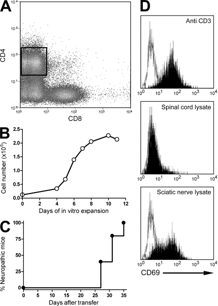
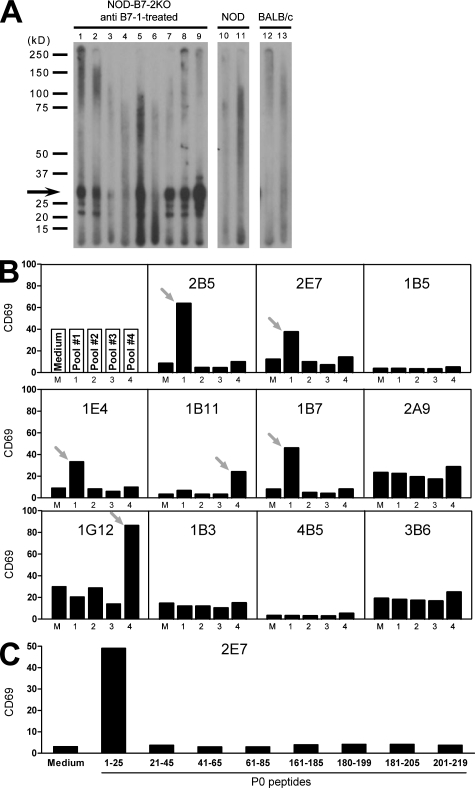
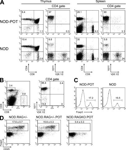
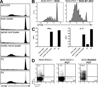
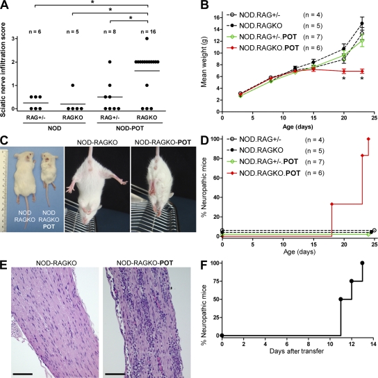
Autoimmune-prone nonobese diabetic mice deficient for B7-2 spontaneously develop an autoimmune peripheral neuropathy mediated by inflammatory CD4(+) T cells that is reminiscent of Guillain-Barré syndrome and chronic inflammatory demyelinating polyneuropathy. To determine the etiology of this disease, CD4(+) T cell hybridomas were generated from inflamed tissue-derived CD4(+) T cells. A majority of T cell hybridomas were specific for myelin protein 0 (P0), which was the principal target of autoantibody responses targeting nerve proteins. To determine whether P0-specific T cell responses were sufficient to mediate disease, we generated a novel myelin P0-specific T cell receptor transgenic (POT) mouse. POT T cells were not tolerized or deleted during thymic development and proliferated in response to P0 in vitro. Importantly, when bred onto a recombination activating gene knockout background, POT mice developed a fulminant form of peripheral neuropathy that affected all mice by weaning age and led to their premature death by 3-5 wk of age. This abrupt disease was associated with the production of interferon gamma by P0-specific T cells and a lack of CD4(+) Foxp3(+) regulatory T cells. Collectively, our data suggest that myelin P0 is a major autoantigen in autoimmune peripheral neuropathy.


Figures

Figure 1.
Figure 1.
Generation of peripheral nerve–specific T cell hybridoma. (A and B) CD4+ T cells were sorted from the nerves of neuropathic NOD-B7-2KO mice (A) and expanded in vitro (B). Data are representative of 15 sorts and expansions. (C) 2–5 × 10⁶ expanded cells were transferred into NOD-SCID or NOD-RAGKO immunodeficient mice, and recipients were followed for the development of neuropathy (n = 5 mice from two independent experiments). (D) CD4+ T cell hybridomas were stimulated with APCs alone (open histogram) or in the presence of the indicated stimulus (shaded histograms), and CD69 up-regulation was assessed. Clone 2E7 is shown. Data are representative of four experiments.
Figure 2.
Figure 2.
Myelin P0 is targeted by the autoimmune response in NOD-B7-2KO mice. (A) Immunoblotting of sciatic nerve extracts with serum from individual anti–B7-1–treated NOD-B7-2KO neuropathic mice (lanes 1–9), nonneuropathic NOD mice (lanes 10 and 11), or BALB/c controls (lanes 12 and 13). Data are representative of five to nine independent experiments for each serum. The predominant 25–30-kD reactivity is indicated with an arrow. (B) Nerve-specific CD4+ T cell hybridomas were examined for reactivity to P0 as in Fig. 1 D, using pools of overlapping peptides spanning the P0 protein. Data are representative of two experiments. Arrows indicate reactive clones to a specific pool of peptide. (C) Activation of the 2E7 hybridoma by individual P0 peptides from pools #1 and #4. Data are representative of two experiments.
Figure 3.
Figure 3.
Phenotypic analysis of POT TCR-Tg mice. (A) Phenotyping of CD4+ and CD8+ T cells in the thymus (left) and spleen (right) of NOD-POT TCR-Tg mice (top) and transgene-negative littermates (bottom). (B) Phenotyping of CD4+ T cells in the spleens of NOD-RAGKO-POT TCR-Tg mice. (C) Foxp3 expression in the spleens of the indicated 12-wk-old mice (histograms are gated on CD4+ T cells). Numbers indicate percentages of Foxp3+ cells in the CD4 population. (A–C). (D) CD25 and Foxp3 expression among CD4+ cells in the spleens of the indicated 19-d-old mice. Percentages ± SD are indicated. Data are representative of two or more experiments.
Figure 4.
Figure 4.
Proliferation and cytokine production of P0-specific NOD-POT T cells. (A) Proliferation of CFSE-labeled NOD-POT T cells in the presence of APCs alone (gray histogram) or APCs and the indicated stimulus (black histograms). Histograms are gated on the CD4+ population. (B) Proliferation of CFSE-labeled NOD-POT spleen cells in NOD or NOD-B7-2KO recipients (22 wk of age) after 4 d (inguinal lymph nodes are shown). Histograms are gated on the Thy1.1+CD4+ population. (C) Production of IFN-γ (left) and IL-17 (right) in the culture supernatant of NOD-POT cells activated by the indicated stimulus. (D) Production of IFN-γ and IL-17 after stimulation of spleen cells from the indicated mice with PMA and ionomycin. Dot plots are gated on CD4+ T cells. Numbers indicate percentages of cells in the quadrants. Data are representative of two or more experiments.
Figure 5.
Figure 5.
Spontaneous development of a fulminant form of neuropathy in NOD-RAGKO-POT mice. (A) Quantification of infiltration in the nerves of indicated mice at 3–4 wk of age. Horizontal bars indicate means. *, P < 0.05. (B) Weight in the indicated mice as a function of age. Results represent cumulated data from two different experiments. Data are means ± SEM. (C) Clasping phenotype in a representative NOD-RAGKO-POT mouse compared with the normal plantar reaction in a NOD-RAGKO littermate, and weight deficit in NOD-RAGKO-POT mice at 3–4 wk of age. (D) Neuropathy in the indicated mice as a function of age. (E) Intense infiltrate in the sciatic nerve of NOD-RAGKO-POT mice. Bars, 250 µm. (F) Adoptive transfer of neuropathy by spleen cells from NOD-RAGKO-POT mice into NOD-SCID recipients (n = 4). Data are representative of two independent transfers. Videos 1–3 are available at http://www.jem.org/cgi/content/full/jem.20082113/DC1.

References

    1. Kieseier B.C., Kiefer R., Gold R., Hemmer B., Willison H.J., Hartung H.P. 2004. Advances in understanding and treatment of immune-mediated disorders of the peripheral nervous system.Muscle Nerve. 30:131–156 - PubMed
    1. Miller S.D., Turley D.M., Podojil J.R. 2007. Antigen-specific tolerance strategies for the prevention and treatment of autoimmune disease.Nat. Rev. Immunol. 7:665–677 - PubMed
    1. Salomon B., Rhee L., Bour-Jordan H., Hsin H., Montag A., Soliven B., Arcella J., Girvin A.M., Miller S.D., Bluestone J.A. 2001. Development of spontaneous autoimmune peripheral polyneuropathy in B7-2–deficient NOD mice.J. Exp. Med. 194:677–684 - PMC - PubMed
    1. Bour-Jordan H., Thompson H.L., Bluestone J.A. 2005. Distinct effector mechanisms in the development of autoimmune neuropathy versus diabetes in nonobese diabetic mice.J. Immunol. 175:5649–5655 - PubMed
    1. Tang Q., Henriksen K.J., Bi M., Finger E.B., Szot G., Ye J., Masteller E.L., McDevitt H., Bonyhadi M., Bluestone J.A. 2004. In vitro–expanded antigen-specific regulatory T cells suppress autoimmune diabetes.J. Exp. Med. 199:1455–1465 - PMC - PubMed

Publication types

MeSH terms